Tag Archives: OCT

Corona Fog

In the past month or so we’ve increased our testing volume after limiting patient visits during a government mandated stay-at-home order during the COVID 19 pandemic. As we’ve ramped back up, we’ve changed our procedures to limit the risk of transmitting the virus. We’ve instituted stringent cleaning protocols, and installed breath shields on some instruments. All clinical personnel now wear PPE (masks, goggles, gloves) and all patients are required to wear masks while in the clinic. We’ve adapted to this “new normal” pretty quickly and our testing volume is almost back to pre-pandemic levels.

Another part of our new normal is a novel clinical finding that seems to be related to the COVID 19 pandemic. Prior to the pandemic, I hadn’t observed this finding. As our numbers increased, I started noticing artifacts that I couldn’t immediately identify in IR fundus images using the Spectralis OCT. I’m wondering how many others in the field have noticed the same thing.

Prominent dark shadow in the infrared fundus image on the left made it challenging to get a good quality OCT image.

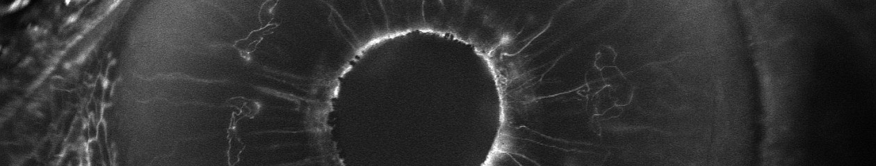
This patient came in two weeks ago. Here you see what appears to be vitreous hemorrhage or debris causing a dark shadow that obscures the view in the IR image. I really struggled to get good images. The OCT was adequate, but the accompanying IR fundus image was poor. Those of you that use the Spectralis know that any structure or pathology that is out of focus will typically appear dark because of the confocal pinhole that blocks scattered light from reaching the sensor. So it’s not uncommon to have a compromised view like that if there are media opacities such as dense cataracts, corneal opacities or vitreous pathology, so I didn’t think much of it at first.  

But then just two patients later I saw a similar artifact. This time it changed in appearance while I was imaging and got progressively worse. I had the patient sit back and re-positioned.

That seemed to improve the view, but within seconds it deteriorated again and got worse. It almost looked as if I were watching a vitreous hemorrhage occur live. I had the patient sit back yet again, waited several seconds, and resumed imaging. The artifact disappeared again!

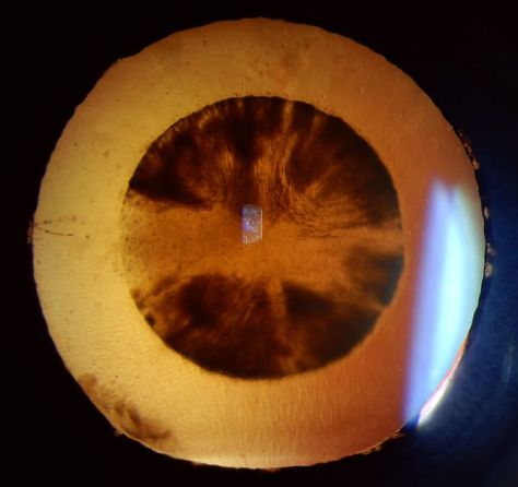
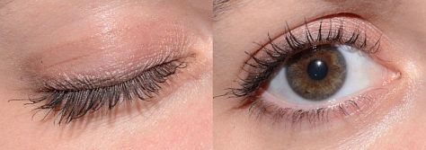
Finally, I looked around the device at the patient’s face to see if there was anything I was missing. And then I noticed it, the patient had an ill-fitting mask and was fogging the lens with her breath! The position of the mask forced her breath directly onto the lens surface of the Spectralis. Because of the confocal nature of the Spectralis, the pinhole aperture causes the artifact to appear dark rather than just a simple fogging seen with a slit lamp, fundus camera, or any other non-confocal device.

I asked a few of my astute imaging colleagues about this and a few had seen it but were initially stumped as well. Collectively we now believe we are seeing a new artifact related to patients wearing masks. One of my colleagues thought it was more prominent with the SPECTRALIS 102 degree wide angle lens. That may be because of the different working distance from the patient (closer).

Shadowing on a Spectralis fluorescein angiogram with 102 wide angle lens from breath fog.
Image courtesy Gary Miller, CRA, OCT-C, FOPS

Looking back I realized that I had struggled with many patients during the peak of the lockdown. We were only seeing urgent/emergent patients at that time and were not routinely dilating patients in the interest of efficiency. So I assumed the darker fundus images were related to working with smaller pupils. It dramatically effects the confocal fundus image, but not always the OCT image because that component of the device is not confocal. The fogging seems to be more common with certain mask types or on days with high humidity.

Now when it occurs, I often just ask the patient to sit back for a few seconds until the fog clears clears and then resume imaging. Or we sometimes tape the top of the mask to the patient’s nose and cheeks to direct their breath down or to the side.

Changing the OCT working distance to the XL setting may reduce the fogging by changing the angle/distance from the patient’s mask.

Another trick is to change the OCT working distance (XL setting) as if you were trying to capture a longer eye. That effectively changes the angle between the patient’s mask and the front element.

My colleague Gary Miller and I have been unsuccessfully trying to come up with a catchy name for this finding. Here are a few of our attempts, but they’re all pretty lame.

Corona Fog
Pandemic haze
Mask mist
COVID condensation
Masquerade artifact

If you have a good one, put it in the comments section.

Here are the current recommendations from Heidelberg on cleaning optical surfaces in their devices. These recommendations seem pretty universal and would likely be similar to those from other manufacturers as well.

Update: A number of people have asked for a document to share whith coworkers. I created a quick pdf adaptation that can be found here.

Tim Bennett, CRA, OCT-C, FOPS

Celestial Bodies – The Eye and Space

First time viewers of ophthalmic images frequently make the observation that the photos look like something from outer space. Especially when reviewing the round orange retinal photos with their eye doctor, patients often comment, “That looks like the planet Mars.”

Every time it happens I get a chuckle out of it. As if we all truly know what the planet Mars really looks like! But to most people, images of the inside of an eye are foreign and amazing. And there does seem to be a little science fiction aspect to both the appearance of the eye when viewed at high magnification, as well as the technology used to capture these amazing images. There are however, several space analogies that really seem to ring true. Among eye-care professionals, the eyeball is routinely referred to as the “globe”.

Macular Star in a patient with cat scratch neuroretinitis

Many clinical findings are named by their appearance rather than an underlying cause, and several conditions have names derived from their similarity in appearance to objects in space: asteroid hyalosis, macular star, star folds, starry sky, astrocytoma, stellate pattern, etc.

Asteroid hyalosis is comprised of calcium soaps suspended in the vitreous cavity behind the iris. Despite their appearance, most patients with this condition are asymptomatic.

In fact, there are enough conditions like this, that I’ve been able to compile them into the Ophthalmic Jeopardy category: Celestial Bodies.

Transillumination of a thinly pigmented iris in a patient with ocular albinism.

Like images from space, there does seem to be an element of wonder and mystery when we peer inside the globe, so in some ways the analogy makes sense.

Cataract

Many ophthalmic images seem reminiscent of photographs from NASA. Or they may stir our imagination or perception of how objects in space might appear.

Lisch nodules on the iris of this patient with neurofibromatosis are reminiscent of peaks, valleys and craters seen in NASA photographs from planets or moons in our solar system.

There are other connections as well. Some of the photographic techniques used by both astronomers and ophthalmic photographers are actually similar. IR capture, interferometry and stereo imaging are common techniques in both fields.  The principles of  rotational stereo imaging can be applied to both subjects. Filters or lasers of different wavelengths are commonly used to enhance visibility of certain features in both subject types.

Most of these analogies between the eye and outer space, are loose associations rather than a direct connection. There is however, at least one eye condition that can be directly associated with a celestial body. Solar retinopathy is a type of photic injury to the retina that is the result of staring at the sun. This condition typically occurs in patients with psychiatric disorders or under the influence of hallucinogenic drugs.

A case of solar retinopathy with a subtle yellow-white foveal lesion with associated early pigmentary changes.

Some scholars believe that early astronomers, especially Gallileo, went blind as the result of solar retinopathy from viewing the sun through a telescope. It’s important to note that this condition can also occur from viewing a solar eclipse without protective eyewear. The upcoming solar eclipse visible in the U.S. on August 21, may cause a spike in cases of solar retinopathy presenting to emergency rooms and eye clinics. The American Academy of Ophthalmology offers some tips for safe viewing of the eclipse.

In recent years, another connection between outer space and vision has been discovered. It turns out that space travel can have some damaging effects on the human eye. Long-term exposure to microgravity can lead to a hyperopic shift in vision from flattening of the globe. This condition is believed to be related to increased intracranial pressure and is sometimes associated with optic disc edema, cotton wool spots and choroidal folds. Optical coherence tomography (OCT) is used to document  changes in thickness of the retinal nerve fiber layer of astronauts before, during, and after space flight.

I took this OCT selfie a few years back when we had a scientist from NASA visiting our clinic while exploring the possibility of putting an OCT on the International Space Station. She wanted to see the Heidelberg Spectralis in clinical use. After demonstrating on several patients, the scientist asked me if I thought it were possible for someone to take an OCT image of themselves. I pivoted the monitor, control panel, and footswitch around so I could operate the OCT from the patient chair and then captured some images of my own retina. I was showing off a little and smugly cautioned the NASA doctor that this was a difficult feat that only an experienced ophthalmic imager could perform. After all, I’ve been doing this for over thirty years. She paused for a moment and then said, “With all due respect, astronauts are some of the smartest and most talented people on earth. They shouldn’t have any difficulty performing OCTs on themselves after a some brief training.” Suddenly I didn’t feel so smug.

A year or so later, the Spectralis arrived at the International Space Station and it looks like she was right. I heard from some colleagues at Heidelberg that the astronauts were given less than 30 minutes of training on the instrument and mastered it quickly!

It’s pretty cool knowing that astronauts are performing ophthalmic imaging on the International Space Station. I wonder if they ever see any resemblance between the eye and celestial bodies?

Disclosure: I have no financial or proprietary interest in the Heidelberg Spectralis.

Here are some links on the condition that’s effecting the vision of astronauts and the use of diagnostic imaging on the space station:

http://www.vision-research.eu/index.php?id=858

https://www.theatlantic.com/science/archive/2017/01/seeing-in-space/513650/

http://www.utsouthwestern.edu/newsroom/news-releases/year-2017/jan/vision-levine.html

https://www.washingtonpost.com/news/speaking-of-science/wp/2016/02/11/cosmic-breakthrough-physicists-detect-gravitational-waves-from-violent-black-hole-merger/

https://www.healio.com/ophthalmology/retina-vitreous/news/print/ocular-surgery-news/%7B5cd3865b-b1db-473a-8800-e3e3b50c6df5%7D/monitoring-long-term-effects-on-vision-in-microgravity-a-priority-for-nasas-future-flight-to-mars


The Value of Maintaining Certification

Along with holidays and festive celebrations, the end of the calendar year often marks the deadline to complete any recertification requirements in order to maintain your professional credentials.  I recently completed my OCT-C (Optical Coherence Tomographer-Certified) recertification in December, and just received my updated certificate in the mail. That will hold me for another three years. At the end of 2017, my other professional credential, the Certified Retinal Angiographer, will be up for recertification. It will mark my 30th year of proudly holding the CRA.

I’m not alone when it comes to maintaining these voluntary certifications. Johnny Justice Jr. continues to set an example by maintaining the CRA credential that he originally obtained in 1979 (the first time it was offered). That’s amazing to me.  Several others from that inaugural group of CRA recipients, including Peter Hay, Phil Chin, Tom Egnatz, and Chuck Etienne, also maintain their CRA after all these years.  None of them have anything to prove at this point in their careers, especially Johnny. He is a pioneer in the profession and was the driving force and founding member of the Ophthalmic Photographers’ Society. He is a well-known author and lecturer, and is universally considered the “Father” of our profession. He certainly doesn’t need the CRA to gain employment, or practice in his chosen profession. He proudly maintains his CRA out of respect for the credential, and what it means to the profession that he helped to create. It clearly has value to him after all these years and all his accomplishments.

In a highly technical field such as ophthalmic imaging it may seem surprising there are no licensure or certification requirements. Certification is strictly voluntary to perform in these roles. It is estimated that less than half the people working as ophthalmic photographers, assistants, or technicians are certified. It’s not easy to obtain certification, and it shouldn’t be. After all it’s meant to identify individuals who have demonstrated a designated level of competence in their field. It takes knowledge, skill, and experience to successfully complete the examination process for certification. Anyone who has completed this process knows the significant effort that is required.

So why get certified if it isn’t required, universally recognized, and takes significant effort? It’s about value. The benefits of certification are often tangible: increased job satisfaction, enhanced job mobility, increased earning power, and a competitive advantage for advancement or the best employment opportunities. But the benefits don’t stop with the certified individual. They also extend to the employer, ophthalmologists, insurers, and most importantly, our patients and their families. All of these groups benefit from knowing they are dealing with a recognized professional.

Certification also helps establish a professional identity and recognition by your peers. It certainly did that for me. In fact, I worked in the field for nearly ten years before initially pursuing certification. I decided it was time to move on in my career and needed certification to gain access to the best jobs. It worked, as my next employer required the CRA as a condition of employment. But I was pleasantly surprised by the additional benefits that certification provided. Although I was very skilled in the technical aspects of photography and had worked at one of the most prestigious institutions in the world, no one outside my place of employment knew my name. That all changed when I obtained my CRA. I suddenly had the respect of my peers and began receiving invitations to play a role in professional activities in the OPS and beyond.  Certification seemed to be “the price of admission” to important networking opportunities that lead to leadership roles in the OPS and JCAHPO. From there came many opportunities to share my knowledge through lectures and publication. And it all started with certification.

But the process doesn’t end when you first receive your credentials. Certification is much more than a one-time achievement. It is a dynamic, career-long commitment to continued education, assessment, and professional development. There is incredible value in attaining, and also maintaining, your certification.

Book Review

Optical Coherence Tomography and OCT Angiography: Clinical Reference And Case Studies

Darrin A. Landry, CRA, OCT-C
Amir H. Kashani, MD, PhD

Bryson Taylor Publishing, 2016
ISBN–‐13:978–‐1523976867
ISBN–‐10:1523976861
www.BrysonTaylorPublishing.com

In the early days of retinal angiography, photographers often worked very closely with ophthalmologists, learning together as they explored the diagnostic uses of fluorescein angiography and unraveled the complexities of interpreting the fascinating images they were capturing. This spirit of scholarly collaboration between imager and physician continues today in a new book: Optical Coherence Tomography and OCT Angiography, Clinical Reference And Case Studies by Darrin Landry and Amir Kashani. These authors are both well respected in their respective fields as educators and authors. Together they have created a timely textbook that will appeal to members of both professions.

Before receiving an advance copy of this book for review, I anticipated that the content would focus almost exclusively on OCT angiography.  I was pleasantly surprised to find that although the book features OCT-A prominently, it is much more than a text on this new state-of-the-art technology. It appropriately places OCT-A in the context of multiple imaging modalities to assist in diagnosis of a variety of retinal conditions.

The authors have produced a book that is part tutorial, part clinical atlas, and a collection of over forty cases that “puts it all together” using multiple imaging modalities with clinical descriptions. The book is divided into three sections:

Section 1. OCT and OCT Angiography

The introductory section will be particularly useful to imagers as it includes a basic overview of OCT and OCT-A technology, followed by a discussion on pattern recognition, normal anatomy and layers of the retina, how to move the scan pattern, recognizing artifacts, EDI/FDI and a basic primer on OCT-A. The OCT-A primer explains how this technology scans through the z-axis and detects motion to identify the retinal vasculature including the deep retinal plexus.  It includes a discussion of artifacts specific to OCT-A . This section will be especially helpful to those new to OCT and OCT-A, and anyone preparing for certification as an OCT-C.

Section 2. Atlas of Images and Disease Pathology

This section is an atlas of retinal OCT findings organized in anatomical order from the vitreous to the choroid. For each condition, the text includes a brief discussion of the disease process, clinical findings, and appearance in multiple modalities. For each condition, there are multiple image examples providing a full spectrum of potential findings for that disease. For instance, there are over twenty different examples of epiretinal membrane. Novice imagers will find this variety especially helpful in learning to recognize different manifestations of a single condition. In addition to common retinal findings the book also includes good examples of less recognized conditions such as outer retinal tubulation (ORT) and reticular pseudodrusen. As expected, retinal vascular diseases include OCT-A examples along with SD-OCT and other imaging modalities including fluorescein and ICG angiography. Experienced imagers will recognize many of these conditions, but the addition of OCT-A will give them another viewpoint and expand their understanding of each disease.

Section 3. Case Studies

The final section of the book is a series of over forty cases where the authors combine a brief medical summary with appropriate imaging modalities for clinical correlation. This format fits well with the current trend of “case-based-learning” in medical education. In many of these cases, OCT-A dovetails nicely with other imaging modalities to increase our understanding of a disease process or help confirm a diagnosis. This quote from the book’s Preface describes the format well “These images are presented in the context of additional imaging modalities to aide the reader in making useful correlations.”

In conclusion, this timely book is well organized and thorough, without becoming unwieldy. It is easy to navigate between sections if you want a quick reference on OCT anatomy or to look for examples of specific retinal conditions and how they may appear on OCT, OCTA and other imaging modalities. With over a thousand images and forty cases, to say that this book is generously illustrated would be an understatement. It is an impressive collaboration between an ophthalmic imager and a retinal specialist that should appeal to a wide audience that would include ophthalmic imagers, retinal technicians, residents in training, and clinicians wanting a reference for clinical correlation between modalities.

From a personal standpoint, I think it’s great to have an ophthalmic imager making a significant contribution to the ophthalmic literature. Darrin’s collaboration with Dr Kashani serves as a model for what imagers can accomplish when we collaborate with physicians on a professional level.  The spirit of collaboration between professions is echoed several times in the book including this statement from the Introduction, “Constant and close communication between the physician and imager is very essential.”

Reviews like this often end with a cliché that suggests that everyone in the profession should “add this book to your collection” or “keep a copy on your bookshelf”. I’ve tried to avoid those clichés, but honestly, I am happy to have this book in my collection and plan to keep it handy in clinic for reference, especially as we integrate OCT-A into our own diagnostic armamentarium.


To Blink, or not to Blink?

It seems almost too obvious to mention, but just like you can’t see through a window when the window shade is pulled down, you cannot view or image the interior of the eye through closed eyelids.

close-open1-640

Obviously we need fully retracted upper and lower lids to get the best view of the fundus with our fundus camera, SLO, or OCT. Because these are noncontact imaging techniques, image quality is also dependent on a regular ocular surface and clear ocular media. An intact tear film is an important optical component of the ocular media. Simply put, to get the best images we need to strike a balance between fully retracted lids and frequent blinking to maintain the tear film.

miller pre-post-640
Top: Shadow from partially retracted upper lid appears at the bottom of the fundus image and degrades the OCT signal. Bottom: fully retracted lid improves the illumination of fundus image and improves signal strength in the OCT.

mccalister pre-post 640
Lashes partially obscure the retinal view in the top image. A fully retracted lid improves the view.

Many patients are nervous about their visual symptoms and what diagnosis the imaging procedure might detect. They often try hard not to blink during the session thinking it will help you get the best images. But their tear film will break up during this time and the view will become compromised until they blink again. And they often apologize for blinking!

To compound this dilemma, these imaging tests are often performed after a patient has undergone an extensive screening workup that includes IOP measurement, and application of topical anesthetic and dilating solutions. Patients may also undergo gonioscopy or macular contact lens examination prior to imaging. A disrupted tear film is an unintended side effect of these procedures and can adversely affect imaging quality.

pre-post blink 640
Top: irregular ocular surface causes degradation of both the fundus image and OCT as the tear film breaks up from lack of normal blinking. Bottom: after a few blinks, the view improves dramatically. Artificial tears would similarly improve the view.

It may seem counter-intuitive, but encouraging patients to blink frequently during imaging sessions can improve cooperation and image quality in fundus photography and OCT imaging. In our clinic, patients are often surprised that we encourage them to blink, having had procedures done in other clinics where they were sternly cautioned against blinking. In my experience as a consultant and workshop instructor, I have often heard OCT operators repeat the words “Don’t blink!” while performing a raster scan pattern that may take several seconds to capture.

cirrus blink2-640
Artifact from blinking during a volume scan. Timing the patient’s blinking pattern can avoid this type of artifact.

They know that a blink will result in an artifact in the volume map, but fail to recognize the need for frequent blinking. I don’t really blame the operator. Often that’s how they were taught to perform the scan during a workshop or training session by the manufacturer’s trainer:

“Don’t blink! Don’t blink! Don’t blink! Don’t blink! Don’t blink! Don’t blink! Don’t blink!….”

No wonder the patients are afraid to blink! Frequent blinking not only refreshes the tear film, it makes the patient feel more comfortable and ultimately more cooperative. You’ll soon learn to recognize a patient’s blinking rhythm and you can time your image capture just as their upper lid is retracting after a blink. Gently encourage the patient by saying, “hold your gaze for just a moment” when you need just a second or two longer to capture a good image. When frequent blinking doesn’t work, application of artificial tears can also make a difference in patients with dry eyes or compromised tear film.

blink clark 4-up-640
Partial versus fully retracted upper eyelid. Image quality is compromised by the lid and lashes in the left images. Gently retracting the upper lid immediately after the patient blinked improves image quality.

During fundus photography, the flash of the camera will cause an involuntary blink that helps refresh the tear film. If the lid or eyelashes obscure the view, gentle retraction of lids with a finger or q-tip may help. You don’t need to forcefully tug on the lid, just retract it a couple of millimeters to get any lashes out of the way and reveal the entire pupil. Patients are often still able to blink with this mild retraction of the upper lid.

So encourage your patients to blink regularly and learn to capture the best images in between the blinks. If it weren’t for all the blinks, anyone could do this job!

“this camera takes great pictures”

When performing fundus photography or angiography, patients often ask about the technology used to help diagnose their ocular condition. After explaining that it’s a form of photography, the conversation will often turn to cameras. Patients sometimes ask for advice on what type of camera to buy for personal use. Often they’ll tell me about a particular camera they have and will invariably say “the camera takes great pictures”.

I’ve always found this expression and the concept behind it quite amusing. It assumes the person looking through the viewfinder and pressing the shutter button has nothing to do with it! On several occasions these conversations took place at a VA hospital while photographing vets. They’ll talk about having purchased a camera overseas, such as a Voigtlander, Zeiss Contax, or Leica in Europe during WWII, or a Nikon while stationed in Japan, Korea, or Vietnam. Without fail, they’ll tell me that “the camera takes great pictures”. All are quality cameras with good optics, but none of them are capable of taking pictures on their own. Someone has to compose the image and press the shutter.

A related misconception has occurred in ophthalmic imaging over the past decade. With all the automated features such as eye tracking, sampling, auto-alignment and  auto-exposure, etc. in the current crop of instruments, there is a perception that retinal imaging is simple and easy to perform. The implication is that the machine takes the picture, not the operator.

screengrab-640

It’s true that recent advancements in technology have improved and simplified image capture, but I still believe that skilled photographers produce the best diagnostic images. I once had a conversation with an overly-enthusiastic OCT salesman who claimed his instrument was better than the competition. As proof, he showed me two images of the same patient taken with two different OCT instruments. “See”, he said, “The competition’s instrument missed the pathology”. I corrected him saying that it wasn’t the instrument that missed the pathology, but the operator.

YurkovicD3 free scan
High resolution line scan of a small retinal arterial macroaneurysm captured using the free scanning technique.

This sparked an interesting debate. I argued that a skilled OCT operator, given appropriate direction from the ordering physician, would not have missed the area of interest. Ultimately we agreed it’s up to the operator to make the best use of the technology they have available.

Several famous photographers have weighed in on the relationship between camera and photographer in producing great images. I love these quotes:

“You don’t take a photograph, you make it.”
Ansel Adams

“The camera doesn’t make a bit of difference. All of them can record what you are seeing. But, you have to SEE”. 
Ernst Haas

“The camera is an instrument that teaches people how to see without a camera.”
Dorothea Lange

 “Photography is the art of observation… I have found it has little to do with the things you see and everything to do with the way you see them.”
Elliot Erwitt

“There is nothing worse than a brilliant image of a fuzzy concept.”
Ansel Adams

And finally, whenever someone states, “This camera takes great pictures”, I reply with another common quote that’s been attributed to several people, including boxing trainer Roger Mayweather, basketball great Charles Barkley, and possibly even Confucius:

“It ain’t the tools, it’s the carpenter”.

Lenz lens3-672
Slit-lamp photograph of dislocated intraocular lens implant and iris erosion shown in transillumination. Slit-lamp imaging relies on advanced photographic lighting techniques to accurately document the condition of the eye.

Voigtlander camera image from Wikimedia Commons: https://en.wikipedia.org/wiki/File:Vitorets.JPG#filelinks

Mystery Diagnosis: Chairman’s Challenge

One of the cool things about being an ophthalmic imager is the teamwork that can develop with the ophthalmologists you work for. They rely heavily on the images you provide to help diagnose various eye conditions. I’ve been fortunate to work with physicians that rely not only on my imaging expertise, but also my experience in recognizing clinical features of unusual conditions. Recently, one of our medical retina specialists (my boss) challenged my diagnostic skills with an unusual case.

challenge sticky
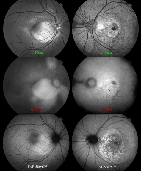
The photo request came through as a post-it note that simply listed the patient’s name and the words: “Fundus photos & OCT”. There was no diagnosis listed or specific instructions given. I called the patient in to the imaging suite and it was a teenage girl accompanied by her mother. I asked if the doctor had given them any paperwork for me and the patient’s mother said, “No. Doctor Q said he wanted to see if you could guess the diagnosis”. Dr. Q and I have worked closely together for twenty years and I can often anticipate exactly what type of images he needs without much direction. But this was unusual. The lack of information was clearly deliberate on his part, which got me thinking:

Is he testing me?

Or does he need my help in making the dx? Yeah that’s it.

Wait… He’s Chairman of the Department, a sub-specialist in medical retina, has written a textbook/atlas of retinal diseases….

He’s clearly testing me, but it must be a rare condition. Hmmm….

Aha! He probably wants images for his new book.

These thoughts are swirling through my mind as I start with an OCT of the right eye. The IR fundus image shows a large oval area that is dark from elevation. It’s suggestive of a serous retinal detachment, but the OCT shows no fluid. The retinal pigment epithelium is pushed forward suggesting a choroidal lesion.

OsteomaA

I start going through a differential diagnosis process in my mind as I continue to capture images:

Nope, not a serous detachment. There’s increased choroidal reflectivity and thickening which suggests a choroidal tumor or nevus of some type. Could it be a melanoma or an osteoma?

Osteomas are pretty rare. I’ve seen one case in the past 30 years. Probably not…

I move to the fellow eye and the OCT shows significant pigmentary changes, subretinal fluid, and what looks like a choroidal neovascular membrane.

OsteomaB

Hmmm…. The findings are more dramatic in the left eye. Looks like maybe an exudative process in a young patient … could it be Coat’s Disease?

I move the patient to the fundus camera and peer through the eyepiece.

osteomaR

Yikes! I wasn’t expecting that. Maybe I was right to think Coat’s.

Coat’s disease is an idiopathic developmental retinal vascular abnormality in children.

coats1

Characteristic findings include telangiectatic vessels with aneurysmal dilation and exudative detachments.

But Coat’s doesn’t quite fit in the right eye. Maybe Vogt Koyanagi Harada disease (VKH)?

VKH1

Features of VKH include: choroidal thickening, hyperemic optic disc, multi-lobed serous detachments and de-pigmentation and clumping of the RPE late in disease.

Although some of the features fit, the characteristic serous detachments of VKH aren’t present. Both Coat’s and VKH are unusual, but not rare. At least not rare enough for Dr. Q to challenge me like this.

I shifted the camera to the left eye to take photos. Once again I was surprised at the clinical appearance.

osteomaL

At that point I must have smiled a little because the patient’s mom asked if I knew the condition. I said yes. Although it looked somewhat like Coat’s, I was almost certain it was a case of bilateral choriodal osteomas. A moment later, Dr. Q came into the imaging suite and confirmed the diagnosis as choroidal osteoma. Sure enough, he wanted good photos for his next book!

osteoma5

Choriodal osteomas are benign ossifying tumors of the choroid composed of mature bone elements. They often demonstrate a thin plaque-like yellow-tan lesion in the macula with sharp, scalloped borders. They are usually unilateral, but can be bilateral. Symptoms include metamorphopsia, scotoma and blurred vision. Vision loss may be due to direct tumor involvement or secondary to choroidal neovascularization with subretinal fluid, lipid, or hemorrhage.

Now that the mystery was over and I had passed the test, Dr Q. handed me the patient’s chart as I finished up the photo session. Her clinical findings were listed:

  • 15 y.o. female
  • VA: 20/25 OD 6/200 OS
  • IOP: 15/10
  • Pupils: Left APD (afferent pupillary defect)
  • SLE: WNL OU
  • DFE:
    • OD: Orange placoid elevated lesion
    • OS: Yellow-orange lesion with well-defined borders. Pigmented CNVM w/ subretinal hemorrhage
  • B-scan ultrasonography demonstrated classic highly reflective plaque-like structures with an acoustically empty region behind the tumors.

osteoma8

This case prompted me to look up the previous case I had encountered nearly twenty years ago. It was before the advent of OCT, but the osteoma was well documented with multi-spectral monochromatic imaging, color fundus photography, fluorescein angiography and B-scan ultrasonography. Dr. Q included that case in his retinal atlas and I had used it lectures on multi-modality imaging. The clinical appearance was quite striking and unforgettable.

osteoma12

It’s great when physicians challenge their staff to take an active role in a team approach to eye care. I’m fortunate to work with physicians like Dr. Q, who challenge me to not only capture high quality images, but to recognize the clinical features of routine and rare cases. Luckily, I passed this test!

The Eye is Like a Camera

“The eye is like a camera.”

I first heard this expression many years ago when a retina specialist was explaining the results of a fluorescein angiogram to a patient and her family. He went on to explain that her retina represented the “film” in the camera and that it was swollen with fluid and wrinkled, which was the cause of her blurred vision. Everyone present easily understood the analogy, as 35mm cameras were commonplace at the time. The idea of warped or wrinkled film resonated as an analogy.    

Although it was my first time hearing this comparison, it was not a new or unique concept. The compelling connection between the eye and camera dates as far back as the 16th century when Leonardo DaVinci drew a comparison between the camera obscura and the human eye. In the next century, Della Porta, Cigoli, and Descartes also made a similar connection in their writings on optics and vision.

With the introduction of practical photographic methods in 1839, the analogy soon extended beyond the optical comparison between eye and camera obscura to include the comparison between vision and imaging. As photography and cameras quickly grew in popularity, scientists, physicians and ophthalmologists such as Helmholtz and LeConte advanced the analogy between photographic camera and the eye.​

“The further explanation of the wonderful mechanism of the eye is best brought out by a comparison with some optical instrument. We select for this purpose the photographic camera. The eye and the camera: the one a masterpiece of Nature’s, the other of man’s work.”

            Joseph LeConte

​From the earliest days of photography, several investigators sought to use the camera to document the condition of the eye. Due to the technical limitations of available photosensitive materials and the difficulty in illuminating the interior of the eye, fundus photography became the “holy grail” of medical imaging. Over the next several decades there were incremental advances in optical instrumentation and photographic processes, all moving steadily towards practical examination and photography of the eye. The most notable of these milestones was the 1851 introduction of the ophthalmoscope by Helmholtz.

Lucien Howe, one of the early pioneers in fundus photography echoed the idea of eye as camera in his 1887 article in which he described having captured the first recognizable fundus photograph:

“We know that the eye itself is a camera, and when placed behind an ophthalmoscope it has pictured upon the retina an image of an eye observed.”

Lucien Howe

The analogy was taken yet a step further by Mann who presented a paper utilizing animal eyes as an actual camera by placing photographic film against an aperture cut in the posterior pole of the globe.

“The main object has been to demonstrate that the eye can actually be used as a camera, a fact which is of interest chiefly because of the frequent comparison between the two.”

William Mann


From that point on there were several milestones in the evolution of both photography and ophthalmic photography that culminated in reflex-free fundus cameras equipped with electronic flash by the 1950’s. The modern fundus camera revolutionized ophthalmic photography and provided the platform for the important development of fluorescein angiography techniques by Novotny and Alvis in 1959. Optical coherence tomography has further revolutionized ophthalmic imaging, but fundus photography and angiography remain vital tools in the diagnosis and treatment of ocular disease.

In today’s wireless and digital age, the analogy of eye as camera may not be as universally accepted as it was a generation ago. Most 35mm cameras had a spring-loaded pressure plate on the film door to ensure the film was held flat and firmly in place. It was easy to envision the concept of a piece of film (or a retina) being wrinkled, torn, or warped.

Today’s digital cameras do not open to reveal the fixed sensor inside so the analogy doesn’t resonate as well. Yet the optical comparison between the eye and camera lens still rings true today, even with sophisticated scanning instruments such as SLO and OCT.

“The eye is like a camera.”…. Since the days of DaVinci, this idea has been echoed countless times by philosophers, artists, ophthalmologists and photographers. As an ophthalmic photographer, I consider myself fortunate to have spent a career photographing the human eye: one camera being used to preserve the capabilities of the other.

“The camera exists because men were intrigued by the function of the eye and wished to be able to reproduce on a permanent record that which the eye enabled the brain to record. How appropriate then that ophthalmology has turned the camera into a valuable tool for recording the structures of the eye.”

R. Hurtes

“The eye is like a camera.”

References

Wade NJ, Finger S. The eye as optical instrument: from camera obscura to Helmholtz’s perspective. Perception 2001; 30:1157-1177

LeConte J. The Eye as an Optical Instrument. from Sight: An Exposition of the Principles of Monocular and Binocular Vision. 1897

Howe L. Photography of the interior of the eye. Trans Amer Ophth Soc. 23:568-71 July 1887

Mann WA. Direct utilization of the eye as a camera. Trans Am Ophthalmol Soc 42:495-508, 1944

Novotny HR, Alvis DL. A method of photographing fluorescence in circulating blood of the human retina. Circulation. 24:82, 1961

Meyer-Schwickerath, G. Ophthalmology and photography. AJO 66:1011, 1968

Bennett TJ. Ophthalmic imaging – an overview and current state of the art. Journal of Biocommunication, 32(2):16-26, 2007.

Bennett TJ. Ophthalmic imaging – an overview and current state of the art, part II. Journal of Biocommunication, 33(1):3-10, 2007.

Hurtes R. Evolution of Ophthalmic Photography. International Ophthalmology Clinics. 1976; 16(2):1-22


Me, Myself, and Eye

Although it may seem like a recent phenomenon, the photographic self-portrait has been with us since the dawn of photography. Perhaps the earliest known “selfie” was taken by Hippolyte Bayard (Portrait of a Drowned Man), a Frenchman who claimed to have invented a photographic process prior to the Daguerrotype. The same can be said of William Henry Fox-Talbot (The Reading Establishment).

In recent years, the photographic self-portrait has exploded in popularity into a global phenomenon, fueled by social media sites such as Facebook, Instagram, Twitter, Snapchat, and others. It is estimated that over one million “selfies” are taken every day. A recent search of Instagram returned over 211 million photos with the hashtag “#selfie”. The term “selfie” is believed to have originated in Australia and has been elevated from internet slang to our common vernacular and even inclusion in several formal English dictionaries. In fact, selfie was Oxford Dictionary’s word of the year for 2013!

Everybody seems to be getting into the act including celebrities, politicians, and even the Pope! Selfies are even popular in space.  Astronauts have shared several spacewalk selfies online, and last year, NASA promoted a global selfie project to celebrate Earth Day. They solicited over 36,000 selfies from around the world and created an interactive composite image that can be viewed on their website.

The selfie is so ubiquitous in today’s pop culture that cell phones and digital cameras often include built-in selfie-friendly apps and features such as extra wide angle lenses, articulating screens or front facing screens that facilitate the selfie pose.  You can also purchase selfie sticks to extend the camera to a better vantage point.  These popular items go well beyond the simple self-timer found on many cameras of yesteryear. Selfies are often purposely self-deprecating, campy, cheesy, or irreverent. They are meant to be spontaneous and fun and are not usually taken very seriously. The selfie craze has even spawned the infamous “duck face” pose.

Despite the fun and seemingly harmless spirit behind them, there is a belief that taking selfies can be a sign of narcissism rather than simple self-expression. There is also some concern it can be addicting and unhealthy. But there is a growing trend in telemedicine where patients can take and forward selfies to their doctors to help diagnose or triage the urgency of their condition.  So maybe there are some legitimate uses for selfies.

As a life-long photographer, I’ve taken my share of selfies over the years. I’ve even attempted a few with the equipment I use for diagnostic ophthalmic photography. Some are goofy, and in the spirit of social media selfies. Some are more practical.

Most ophthalmic imaging devices are not what you’d normally consider selfie friendly, at least not in terms of taking a photo of one’s own eyes. Because the optics of these devices are designed for photographing curved surfaces found in the interior of the eye, they usually create distortion when backed up an appropriate distance to take a facial portrait. The effect of this distortion eliminates the need for a goofy facial expression if your goal is just to post a unique selfie on the internet!

But what about useful diagnostic or artistic photos of your own eyes, photos that go beyond distorted face selfies? It’s not only possible, but surprisingly good images can be obtained with some devices.  Non-mydriatic instruments with a monitor that can be pivoted toward the patient/photographer lend themselves to self-imaging, while those with an optical viewfinder (fundus camera) or fixed monitor position (Cirrus) do not. I’ve been able to obtain eye selfies with the Zeiss Stratus, Heidelberg HRT, Heidelberg HRA/OCT, Clarity RetCam, Tomey specular microscope and various handheld external cameras. But, you might ask, “So what?” or “Why?”

Well, there have been times when I needed to check a device during maintenance or a software upgrade and it was convenient to use myself as the patient. Sometimes while training staff to use a device, I’ll demonstrate the procedure on myself. Other times, I’ve needed a quick example of a “normal” eye for a lecture like those above.

Surprisingly, the ability to take eye “selfies” has helped me identify and track pathology in my own eyes. Two years ago I suffered an idiopathic retinal tear with avulsed bridging vessel and persistent vitreous hemorrhage. This was successfully treated with vitrectomy. Like many patients, I developed a cataract after the vitrectomy. I also began to notice some distortion that corresponded to progression of an epiretinal membrane (ERM) in the same eye.

Any time I noticed a change in vision I would repeat an OCT on myself. Over the course of six months I tracked an increase in thickness of about 100 microns. The cataract also progressed and I was scheduled for cataract surgery. Two weeks prior to surgery I noticed a very subtle change in vision and sat down at the OCT like I’d done several times in the past. The OCT detected some cystoid macular edema (CME) from the ERM. Picking up the CME prior to cataract surgery was very beneficial. Preexisting CME can be exacerbated by cataract surgery, so my surgeon began a course of treatment that reduced the edema. My OCT selfies likely helped us avoid more severe or persistent edema by catching it in advance.

Cataract surgery went as planned, but within a few hours of my procedure I began to notice a new visual abnormality: a paracentral gray scotoma. Upon arriving at the clinic the next day for my post-operative check, I immediately did an SLO/OCT selfie and identified an unusual finding that corresponded directly to the scotoma.

SD-OCT demonstrated an area of hyper-reflectivity in the middle retinal layers just temporal to the fovea (green arrows) and the IR reflectance image showed a distinct dark gray lesion. Fortunately, the scotoma began to fade within a few days and so did the lesion. The jury is still out on the exact cause of the lesion but the selfies have enabled us to track improvement of my condition and possibly publish a case report. We believe it may be a case of paracentral acute middle maculopathy (PAMM), a recently described variant of acute macular neuroretinopathy (AMN). It’s rare enough, that I was able to present it at the OPS Rare Case Symposium in Ann Arbor.

As you can see, image quality can be quite good with a little practice. So good in fact that I’ve received a bill from my institution for OCT images that I’ve performed on myself! Here is a double selfie video of a Spectralis IR fundus image showing how easy it is to capture my own epiretinal membrane.

I’m beginning to think maybe I should stop taking selfies of my own eyes. After all I keep finding abnormalities! But there is a growing trend in telemedicine where patients can take and forward selfies to their doctors to help diagnose or triage the urgency of their condition.

Ophthalmic Photographers taking diagnostic selfies: obsessive, silly, or beneficial?