
Introduction
Monochromatic fundus photography is the practice of imaging the ocular fundus with the use of colored or monochromatic illumination. In 1925, Vogt described the use of green light to enhance the visual contrast of anatomical details of the fundus and coined the term “red-free”. The technique is still commonly used today in combination with fundus photography.
Basic Principles
Monochromatic fundus photography is based on two basic principles of light and photography: the use of contrast filters to alter subject tones in black-and-white photographs, and the increased scattering of light at shorter wavelengths. By limiting the spectral range of the illuminating source, the visibility of various fundus structures can be enhanced. This technique is most effective when combined with high-resolution black-and-white film or monochrome digital sensors.
Contrast filters adjust the monochromatic tonal rendition of different colors by introducing brightness differences between colors that would ordinarily reproduce as similar tones of gray. Commercial photographers have used contrast filters for years to adjust the tonal rendition of portraits and landscape photos, often making them appear more dramatic. A subject color will appear lighter when photographed through a filter of the same color, and darker when photographed through a filter of its’ complementary or opposite color. For example, a red object would appear lighter if exposed through a red filter; while the same red object would appear darker when photographed through a cyan filter.
The visible spectrum can be divided into thirds; the short, intermediate, and long wavelengths, which represent the primary colors: blue, green, and red. The following series of photographs demonstrates the effect of the three primary color filters on an outdoor scene of a barn.

Red, green and blue filters each transmit one-third of white light while blocking the other two-thirds. The red filter lightens the red barn while darkening the grass, foliage and sky. The blue and green filters have similar effects – lightening objects that are the same color as the filter while darkening the other two primary colors.

Landscape photography can also be used as an analogy to demonstrate the increased scatter of short wavelength radiation. Ultraviolet and blue wavelengths sometimes have an adverse effect on photographs of distant outdoor scenes. Short wavelength radiation is scattered more sharply than long wavelengths by small particles in the atmosphere. This phenomenon is known as Rayleigh scattering. When there is an increase in atmospheric particles, short wavelength scattering causes the appearance of haze. Landscape photographers traditionally use yellow or red long-wavelength filters to reduce atmospheric haze with panchromatic black-and-white films. “UV” or “Skylight” filters are commonly used to eliminate ultraviolet and short-blue radiation to correct the bluish cast on daylight balanced color film in photographs of mountains, snow scenes, water or open shade due to unwanted short-wavelength scatter. In monochromatic fundus photography we sometimes welcome the scattering of short wavelengths. Scatter can increase visibility of the semi-transparent anterior retinal layers.

The next series of photographs demonstrates the monochromatic principles and their effect on fundus photographs.

The same three primary color filters used for the previous demonstration have been applied to images of the fundus. Various fundus structures become more or less visible with each distinct monochromatic rendition.

Wavelengths
Blue light increases visibility of the anterior retinal layers, which normally are almost transparent in white light. Blue light is absorbed by retinal pigmentation and blood vessels, providing a dark background against which specular reflections and scattering in the anterior layers of the fundus is enhanced. The retinal nerve fiber layer, the internal limiting membrane, retinal folds, cysts and epiretinal membranes are examples of semi-transparent scattering structures that are enhanced with short wavelength illumination. Because of excessive scattering at very short blue wavelengths, fluorescein exciter filters at blue-green wavelengths of 490 nm are often used. Cataracts, corneal edema or any other cause of scattering in the ocular media, can limit the effectiveness of these wavelengths. When light is scattered before it reaches the retina, the view can be quite hazy at shorter wavelengths. The effectiveness of blue or blue-green contrast filters may be severely reduced if patients are routinely photographed after a complete examination that includes applanation tonometry and gonioscopy. These two procedures often compromise the clarity of an otherwise clear cornea. Any residual topical fluorescein from corneal examination or tonometry will fluoresce under blue illumination, causing a reduction of contrast during photography. It may seem impractical, but it is strongly recommended that monochromatic photography be done before these exams or on a separate visit.

Green light is also absorbed by blood, but is partially reflected by the retinal pigmentation in comparison to blue light. There is less scatter than with shorter wavelengths, so media opacities are not quite as troublesome. Green light provides excellent contrast and the best overall view of the fundus. Because the spectral sensitivity of the human eye peaks in the green – yellow portion of the spectrum, 540-570nm green filters are easy to view and focus through. Green light enhances the visibility of the retinal vasculature, and common findings such as hemorrhages, drusen and exudates. For this reason, green filter “red-free” photos are routinely taken as baseline images before fluorescein angiography.

Retinal pigmentation appears progressively lighter and more transparent in red light, revealing more of the choroidal pattern. Red filters available for fundus cameras usually peak at 625-640nm. Overall fundus contrast is greatly reduced with red illumination as many retinal structures are red in color. Retinal vessels look lighter and less obvious at longer wavelengths, with the oxygen-rich arterioles appearing very light in comparison to the venules. The optic nerve also appears very light and almost featureless. Red light is useful for imaging pigmentary disturbances, choroidal ruptures, choroidal nevi and choroidal melanomas.

Filters
A variety of filter types can be used for monochromatic fundus photography. Absorption filters, which are the most common filter type for general photographic use, consist of organic dyes coated on an acetate or glass substrate. They selectively absorb certain wavelengths, while transmitting others. The spectral transmission curves of absorption filters exhibit gradually sloped gradients and broad bandwidth. Kodak Wratten gelatin filters, or equivalent, are readily available in many different colors and can be easily cut to fit the aperture of fundus camera filter holders. With heavy use, these filters can become warped or scratched. Glass absorption filters are more durable than gel or acetate filters, but they must be ordered specifically for each model of fundus camera to ensure they fit in the filter aperture. Availability of glass filters in the correct sizes may be limited to just a few color choices.
Interference filters rely on multiple thin coatings of materials with known refractive indices to create ‘interference’ between layers, resulting in rejection of specific wavelengths. Interference filters offer efficient and precise light transmission capabilities with more linear spectral transmission gradients. Narrow-band interference filters are necessary for modern retinal angiography, but aren’t essential for monochromatic photography. Interference filters must be properly positioned so that they are perpendicular to the light path, otherwise the transmission characteristics may be altered.
Filters with peak wavelengths of 490, 540-570, and 615 seem to be the most common choices for monochromatic fundus photography. The fluorescein exciter filter, with peak wavelength of 490nm, can be used as a blue-green monochromatic filter. It is an interference filter designed with a narrow bandwidth that will require an increase in flash illumination compared to other monochromatic filters. Many photographers use the standard green, “red free” filter that was supplied with their fundus camera. Traditionally, the standard green filter is a broadband absorption filter with maximum transmission at 540nm. Some cameras come equipped with a longer-wavelength green filter at 570nm. With the recent advent of fundus autofluorescence imaging, some cameras may have a 580nm yellow-green exciter filter that can also be used for monochromatic imaging. Red filters may be available on some cameras, especially those equipped for ICG angiography.
Common Filters and Wavelengths:
| Color | Peak Wavelength | Wratten Equivalent | Fundus Camera Standard |
| Blue | 450nm | 47B | —- |
| Cyan | 490nm | 44A | Fluorescein Exciter |
| Green | 540nm | 58 | Green “red-free” |
| Yellow-Green | 580nm | —- | FAF Exciter |
| Red | 615nm | 25 | ICG baseline red |
| Red | 630nm | 29 | ICG baseline red |
Film/Sensors
Monochromatic photography traditionally required the use of high-resolution black-and-white films and customized film-processing techniques to maximize diagnostic information. These film-and-developer combinations could be difficult to control, which may explain why monochromatic photography never gained universal acceptance beyond the routine use of baseline red-free photos prior to angiography. Many of the films that were traditionally used for monochromatic imaging such as AgfaPan 25, Kodak Tech Pan, Plus-X and Panatomic X have not been available for many years. Medium-speed films such as Fuji Neopan 100, Ilford Pan-F(50), and Kodak T-Max 100 are still available as of this writing (2008). Efke KB 25, KB 50 and Macophot 25, 100, are low-to-medium speed, high-resolution films manufactured in Europe for a niche market of film traditionalists. These films may be a viable replacement for some of the previously discontinued low-speed offerings. Practically speaking however, these films would still be subject to the traditional photographic hurdles of controlling exposure, contrast, and processing.
Digital Imaging
Digital imaging offers some advantages over film-based monochromatic photography. It facilitates immediate adjustment of exposure settings and easy contrast enhancement, making the monochromatic technique more practical for widespread use. The biggest disadvantage to digital imaging is the linear response and narrow exposure latitude of currently available digital sensors. Proper exposure is essential. Even the slightest amount of over or under exposure can be detrimental to digital image quality. Exposure control requires a delicate balance between flash output, sensor gain and gamma settings. With the instant feedback provided on the monitor, the imager can quickly judge image quality and make necessary corrections to mitigate the limited dynamic range of digital sensors. Note improvement in detail of the optic nerve when exposure and contrast are balanced with gain, flash and gamma settings during capture:

The most practical way to capture digital monochromatic images is with a monochrome sensor that produces grayscale images. This method is analogous to using black-and-white film. Monochrome sensors are more light-sensitive than color sensors, and they maintain higher resolution because all pixels can be exposed to any color of monochromatic light. In addition to this method there are several other ways to obtain monochromatic images using digital technology. Many digital fundus-imaging systems use a single full-color sensor for both color and grayscale imaging. The color sensor can be set for monochrome capture in the same manner that is used for fluorescein angiography with many systems. It should be noted however, that color sensors rely on a mosaic array of microfilters to limit the light sensitivity of each pixel to one of the individual color channels, red, green, or blue, causing a reduction in both light-sensitivity and resolution when monochromatic light is used. Color sensors can also be left in full-color capture mode to record a color image though the monochromatic filter. Post-capture conversion to a grayscale image can be done in the imaging software or in a separate image-editing program such as Photoshop.


Another alternative would be to take full-color fundus photos and then use software to split the color image into the red, green, and blue color channels. This is a remarkably simple way to obtain monochromatic renderings from any full color image.

A disadvantage to this method is the loss of resolution that occurs when viewing just a single channel. It also limits the available monochromatic information to just the three primary colors, red, green, and blue, thereby eliminating the possibility of capturing valuable blue-green renditions of anterior scattering structures such as this example of the same retina through the fluorescein exciter filter at 490nm.

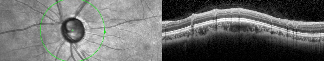
High-resolution digital sensors in combination with monochromatic filters can improve the visibility or detection of many structures that were traditionally imaged with fluorescein angiography, or more recently with optical coherence tomography. Monochrome sensors of three megapixels or better, and color sensors with at least six megapixels of resolution are capable of identifying cystoid macular edema, macular holes, epiretinal membranes and other pathology that may not be obvious on examination or in color photos. High-resolution monochromatic imaging may be useful in cases when OCT imaging is not readily available or as an adjunct to OCT as shown in this example of CME.


Technique
Exposure often needs to be increased to compensate for light loss when the filter is introduced into the light path of the fundus camera. The amount of exposure increase is dependent on the transmission characteristics of the contrast filter and the spectral sensitivity of the specific panchromatic film or digital sensor being used. Pupils should be maximally dilated to maximize light transmission. Photography should be done before gonioscopy, contact lens examination, or applanation tonometry, to avoid excessive scattering at shorter wavelengths. Camera optics must be kept clean for the same reason. Focus should be targeted to the specific retinal level enhanced by the contrast filter in use. For example with a blue-green filter, focus should be on the anterior surface of the retina. Green light is typically focused a little deeper, usually on the retinal vessels, while red light is focused at the level of the RPE or choroid. It is always important to maintain even illumination and proper camera-to-subject distance in fundus photography, but even more so in monochromatic photography. The increased contrast and scattering induced by monochromatic filters can exaggerate any flaws in illumination and alignment. The sequential stereo technique can be combined with monochromatic photography to add additional diagnostic information.
Monochromatic techniques and filter choices are based on the premise that the human fundus is mostly red in appearance, hence the use of “red free” light to darken the deep layers of the retina and choroid. But there is quite a bit of variability in normal fundus pigmentation, much like the normal variation in skin pigmentation in the general population. This makes it difficult to choose contrast filters that will work equally well on all patients. Similar limitations have been noted in the use of contrast filters in general photographic applications. Subject colors are often comprised of too wide a range of wavelengths for filters to be as effective as we would like. Normal variations in fundus pigmentation and the clarity of the ocular media are factors that can lead to inconsistent or disappointing results. Because of these factors, a single monochromatic rendition doesn’t always tell the entire diagnostic story. Technique could be standardized to include the routine use of more than one filter on all patients. Using multiple filters in a photo essay style approach can provide the clinician with more than one monochromatic interpretation of the fundus, such as in this example of an epiretinal membrane with macular pigmentary changes shown at 490nm, 540nm, and 615nm.


