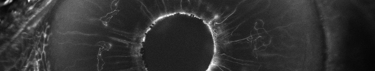

The ocular fundus represents the most important subject in ophthalmic imaging. The fundus is the inside back wall of the eye and includes the retina, optic nerve head and choroid. Fundus imaging presents some interesting challenges. Our goal is to photograph anatomic structures that can be measured in microns, with enough detail to make diagnostic decisions. All this must be done through the pupil, an opening in the iris just a few millimeters in diameter.

Imagine peering through a keyhole into a darkened, curved room that’s a little over an inch in length. In order to visualize the opposite wall of this small chamber, we must illuminate it by shining light through the same small opening we are peering through. This challenge requires specialized optical instruments such as the fundus camera and the confocal scanning laser ophthalmoscope.
In addition to full color photo-documentation of the clinical appearance of the fundus, different wavelength filters or lasers can be used to detect diagnostic features not visible on examination.
Techniques include: