There are three major steps to the digital imaging process: image capture, image processing and image output:
- Capture: Digital image capture describes the electronic recording of light information and its conversion to a set of numbers, which are stored in an image file.
- Processing: Once an image is captured and stored on a computer as numeric data, it can then be processed. Image processing uses computer software to manipulate the set of numbers that were created in the capture process to enhance the original image by adjusting contrast, exposure, color balance, cropping and sharpness. Processing prepares the image for output.
- Output: Image output involves converting the processed numeric data back into a viewable form for display, either onscreen or as printed hard copy. Screen output can take many forms, including computer monitor, remote display, email, LCD projection, and web page display.
Digital images are captured by an array of photosensitive imaging sensors in a camera or scanner. Common imaging arrays include charge-coupled devices (CCD) or complementary metal oxide semiconductor (CMOS) sensors or “chips”. Both sensor types are constructed of an array of photodetectors arranged in a grid or mosaic pattern of pixels. Each detector can capture only one color – red, green, or blue. Scanners usea linear sensor, while cameras incorporate an area sensor.

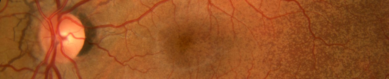
Area sensors are a square or rectangular matrix of photosensitive pixels arranged in rows and columns. They are capable of capturing an entire image at once, which makes them suitable for moving subjects or flash photography. Area sensors are used in cameras like the ones we use for fundus photography or incorporated in DSLR cameras.
Once acquired, ophthalmic digital images can usually be enhanced, printed, archived, retrieved and exported using the proprietary imaging application supplied with the digital system. Images from a photographic session are available instantly and are typically displayed in a proofsheet or thumbnail image format.

Images can then be viewed or printed individually, or in groups (including a “4-up” or “9-up”). Commercially produced fundus imaging systems may include measurement tools calibrated to the magnification of the fundus camera to provide accurate tracking of lesion size.

The enhancement tools supplied with these systems will allow changes to brightness, contrast, sharpness, and color balance to images being displayed or printed. For medical record purposes, it is vital that we retain the integrity of the original image. This is an important issue because digital images can be easily modified. It is important that the original file is not overwritten when enhancements are made. Enhanced images can be added to the patient’s record, but the original image files are stored without modification.