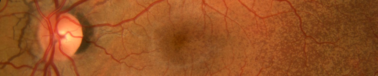

External Ocular Photography
Perhaps the most obvious but least specialized type of ophthalmic photography is what is termed “external photography”. Conventional macro photography equipment and techniques are used to document the external, rather than internal, appearance of the eyes and surrounding lid and facial structures. It is commonly used to document lesions of the eye or surrounding tissues, demonstrate facial nerve anomalies, and record pre and post surgical alignment of the eyes or eyelids.

Point and shoot digital cameras or smart phones are sometimes used for external photography but they are a poor choice because of the typical wide-angle lens, the inability to preset the magnification for repeatable results and the lack of appropriate onboard lighting.
Digital single-lens-reflex (DSLR) cameras are the best tool for high-quality external photography. DSLRs avoid parallax between lens and viewfinder and they offer a variety of compatible lens and electronic flash choices. Magnification for routine external ocular photography ranges from full-face up to life-size 1X. Short telephoto macro lenses are the best choice to provide this range and facilitate repeatable magnification settings. They maintain a natural perspective and a comfortable working distance.
Auxiliary electronic flash units with a variable positioning system provide directional lighting that can be adjusted tangentially to demonstrate texture and elevation or moved to an axial position to illuminate a cavity or to accurately render skin color with even lighting.

In addition to the inherent advantages of using an SLR for magnification and composition, the LCD screen provides instant feedback on the quality and position of lighting, eliminating the need for a continuous modeling light. Photographers can easily view the results and adjust the flash position and intensity to obtain the desired lighting effects based on the specific subject or pathology.

Motility photographs are a series of external photographs showing both eyes together in various positions of gaze. They are used to demonstrate alignment or misalignment of the eyes due to imbalanced extra-ocular muscles or other disorders that can obstruct or limit normal eye movement. In addition to still images these eye movement disorders are sometimes documented with video.
Slit-lamp Photography
Although external photography can capture views of the conjunctiva, cornea and iris with magnifications of up to 2x life size, most high magnification photography of the anterior segment of the eye is done with the use of a photo slit-lamp. The photo slit-lamp is a variation of the slit-lamp biomicroscope routinely used by eye care professionals to light and examine structures in the eye at magnifications up to 40x.
Slit-lamp photography or as it’s also known, photo slit-lamp biomicrography, offers the best opportunity for ophthalmic photographers to demonstrate their photographic skills and creativity. It is a technically and aesthetically challenging discipline that requires a good understanding of composition, magnification, lighting principles and precise exposure control.

The slit-lamp is a horizontally mounted binocular microscope with a specialized lighting system that allows visualization of the transparent and translucent structures in the eye. It derives its name from the slit illuminator that applies the principles of Koehler illumination to an optical condensing system to deliver an adjustable beam of light that is focused at the same plane as the microscope. The slit illuminator and the biomicroscope pivot on the same axis to maintain a parfocal relationship.

Photo slit-lamps use beamsplitters to direct image-forming light to a camera mounted on the instrument, or in some cases two cameras to provide stereo capture.

Electronic flash illumination is delivered through the same condensing optics as the tungsten slit illuminator. Magnification at the film plane is adjustable from approximately 0.7x up to around 5x, depending on the specific instrumentation. The slit beam can be precisely controlled and adjusted from a round or wide rectangular beam down to a very thin slit. One could think of the slit illuminator as a mini studio light with an aperture that can be widely adjusted to limit or direct light output like barn doors on studio lights. A secondary fill light is often available to add background illumination and adjust lighting ratios.

Slit-lamp photography is a complex task. It often requires a series of static images to simulate the cumulative effect of a dynamic slit-lamp examination that is accomplished by panning the microscope across the cornea while simultaneously focusing through various structures and swinging the slit illuminator to highlight areas of interest. Still images must be focused at each layer of interest and care must be taken to eliminate flare or reflections that would normally be ignored in the course of routine examination. It’s useful to approach it as a photo essay. You are trying to tell a story with a series of photos.

Lighting techniques fall into two basic categories, direct and indirect. Direct focal illumination can be delivered axially, or tangentially to produce highlights and shadow. Specular reflection can be used to detect surface irregularities in highly reflective structures. A very thin focal beam is often employed to create a narrow cross-sectional view or “optical section” of the normally transparent cornea and lens.

Indirect illumination can take on many forms. Iris transillumination, sclerotic scatter, and retroillumination of the cornea or lens are lighting techniques that are accomplished by bouncing or scattering light off other anatomical structures such as the iris, retina, and sclera.

Vital stains may be used as an adjunct to slit lighting techniques to enhance visibility of certain ocular surface disorders. Topical applications of rose bengal, lissamine green, or fluorescein can be applied to stain areas of missing or devitalized cells in the cornea and conjunctiva. These dyes are best photographed using diffuse or broad beam, focal illumination. Fluorescein staining can be illuminated with white light to demonstrate yellow staining or with blue light to cause excitation and fluorescence of the dye-stained areas.
Gonio Photography
Auxiliary lenses are sometimes used in concert with a photo slit-lamp to photograph inner structures of the eye that cannot be directly visualized with the slit-lamp. The most common lens type is the gonio contact lens that utilizes internal mirrors angled at approximately 60° to provide observation of the filtering angle of the anterior chamber. The anterior chamber is the space between the cornea and iris, and the filtering angle is the junction of the peripheral cornea, peripheral iris, and sclera. Examination of the angle is important in the detection of structural abnormalities that can contribute to progression of glaucoma.

Gonio photography is the most complex and difficult photographic procedure in ophthalmology. Methylcellulose is used as an optical medium between the contact lens and cornea after the eye has been anesthetized. Bubbles in the methylcellulose can interfere with photographic composition. Stray reflections from the mirror or front surface of the contact lens can cause flare and unwanted artifacts. Given the technical challenges associated with this technique, it is not performed on a routine basis by most ophthalmic photographers.
Specular Microscopy
Specular microscopy is a variation of slit lamp photography that utilizes specialized single-purpose instruments to image the inner surface of the cornea. The corneal endothelium is comprised of a single layer of flat transparent cells that are responsible for pumping fluid out of the cornea to maintain its transparency.

High magnification and specular reflection is necessary to delineate the cell borders to detect cell density and morphology, which are indicators of corneal health. Magnifications range from 20x to 200x. Traditionally, specular microscopes required contact with the patients’ cornea. The latest instruments utilize non-contact microscopy with semi-automated digital analysis of cell counts and morphology.
