In the 1960’s, a number of technological advances in photography helped to usher in a new era of diagnostic testing in ophthalmology, and with it a new profession was born. Ophthalmic photography is a diagnostic discipline that utilizes a number of photographic techniques to help document and diagnose various diseases of the eye.
The photographers that formed the vanguard of this new profession found a shared need to exchange information, collaborate on new techniques, and set standards of practice in the profession. They formed the Ophthalmic Photographers’ Society, Inc (OPS) to fill these needs. Through the collaborative efforts of OPS members, the profession has grown and flourished. Today, OPS members continue to promote and elevate the profession through publication, education, and certification.
Changing health care delivery models and new technological developments have altered the shape of the profession in recent years. Many photographers have expanded their job responsibilities and developed new skills in related fields. As new diagnostic imaging and treatment modalities are developed, the role of the ophthalmic imager will evolve and continue to play an important role in the preservation of sight.
Beginnings of a Profession
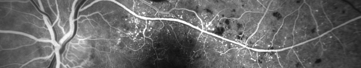
Although there are published accounts of ophthalmic photographic techniques going back to the late 1800s, ophthalmic photography as a profession didn’t evolve until the 1960’s. A number of historical events coincided to provide the foundation for practical retinal photography. Kodak Tri-X film was introduced in 1954. The following year, the Zeiss-Littman modern fundus camera was introduced. It was the first commercially produced fundus camera to incorporate electronic flash illumination.1 In 1959, two medical students, Harold Novotny and David Alvis started a research project to develop a photographic technique to estimate blood oxygen concentrations in the retinal vasculature as a visible segment of the cerebral circulation. In doing so, they worked out the basic techniques of fluorescein angiography and performed the first successful angiogram of a human retina (Figure 1). They reported their results in a landmark paper in 1961.2
Figure 1. Photographs from the first successful fluorescein angiogram in a human subject by Novotny and Alvis. (Image courtesy of Tim Steffens, CRA ,and the Indiana University Medical Center)
Following their report, popularity of the technique exploded, as several investigators studied various clinical applications of angiography, quickly establishing the intrinsic value of the technique.3 Up until this point, most ophthalmic photography had been performed by physicians, researchers, assistants, and in some cases, medical photographers. The sudden enthusiasm for fluorescein angiography created an immediate need for skilled full-time retinal angiographers, providing the foundation for the profession as we know it today.
The first generation of “ophthalmic photographers” to enter the field were mostly medical photographers who already had some experience in fundus photography and were able to make a quick transition to this new subspecialty.4 These early practitioners worked side-by-side with ophthalmologists and retinal specialists in exploring the diagnostic uses of fluorescein angiography and learning together as they unraveled the complexities of interpreting the fascinating images they were capturing. This close clinical collaboration with physicians quickly elevated the profession. Many ophthalmic photographers contributed significantly to the ophthalmic literature of the time and were held in high regard as professional colleagues in ophthalmology. This spirit of scholarly collaboration between photographer and physician continues today in many academic practice settings.
Over time, there were gradual shifts in the professional experience of individuals entering the field. A second generation of fluorescein angiographers came from a cross-section of commercial, industrial, and scientific photography backgrounds, adapting their existing photographic skills to ophthalmic subjects. This was followed by another group of personnel that had experience in ophthalmology as ophthalmic technicians, but with no technical photographic training. The diversity of professional backgrounds of individuals entering the field created a universal need for education and sharing of information. With no real formal training available in ophthalmic photography, practitioners had to learn many necessary skills on the job. Although at least one current biomedical photography degree program offers elective courses in ophthalmic photography, the vast majority of practitioners in the field have no formal training in ophthalmic photography.
In the absence of formal education in ophthalmic photography, it may seem incongruous that the profession provides a high level of academic pursuit and achievement for many members. A number of ophthalmic photographers contribute to the profession of ophthalmology as authors of ophthalmic textbooks and scientific articles, some participate as investigators or sub-investigators in clinical research, and many play an integral role as instructors in ophthalmology residency training programs. Through these contributions and academic achievements, some ophthalmic photographers have even received academic appointments as professors of ophthalmology.
Ophthalmic Photographers’ Society
In the decade that followed the introduction of fluorescein angiography, some of the pioneers in the field occasionally crossed paths while attending ophthalmology meetings. In such a young profession, workers in the field found a great desire for interaction with fellow professionals to share experiences and techniques. In April of 1969, a group of photographers had an informal gathering during the Association for Research in Vision and Ophthalmology meeting in Sarasota, Florida to discuss forming a professional society.5
Founding Members of the Ophthalmic Photographers’ Society Inc
Lee Allen
Yvonne L. Magli
Earl A. Choromokos
Mary T. Mannella
Ogden Frazier
Terrance L. Tomer
Johnny Justice, Jr
Anna Wiley
Roger Lancaster
Don Wong
They agreed to have their first formal meeting in Chicago during the American Academy of Ophthalmology meeting later that year. Attending the first meeting were ten ophthalmic photographers, who set organizational goals, selected interim officers and chose a name (Figure 2). The Ophthalmic Photographers Society, Inc (OPS) was incorporated in July of 1970.
Figure 2. The early days of the Ophthalmic Photographers’ Society. Johnny Justice Jr. CRA, FOPS, principal founding member of the Society, addresses the second organizational meeting of the OPS. Lee Allen, FOPS first OPS President (center) and others look on. March 6, 1970. (Image courtesy of the OPS Historical Archives).
The OPS is a non-profit organization that today counts over 1000 members from 27 countries. Membership is open to anyone with an interest in ophthalmic photography and is made up of photographers, technicians, physicians, scientists, vendors and students. The main objectives of the Society are to provide primary and continuing education in the field of ophthalmic photography, to set and maintain professional standards through certification, and to promote scientific advancement in imaging technology and techniques.
Since inception, the OPS has provided a central forum for the exchange of information through a number of programs and publications. The Society sponsors national and regional educational meetings, offers educational scholarships, publishes the peer-reviewed Journal of Ophthalmic Photography (Figure 3) as well as a member newsletter, maintains a website filled with important news and technical information, and offers certification in ophthalmic photography. All of these programs and member benefits are accomplished almost entirely through the efforts of dedicated volunteers from the OPS ranks, along with generous philanthropic support from our sustaining members.
Figure 3. The Journal of Ophthalmic Photography, a peer-reviewed publication of the Ophthalmic Photographers’ Society. Beth Ann Benetz, CRA, FOPS, Editor.
Despite the highly specialized nature of the profession, the OPS has found shared interests and collaborative opportunities with other organizations, such as ICE, JCAHPO, ICOP, and others. The OPS has been a member of the Institute for Credentialing Excellence (ICE – formerly NOCA ) since 1989. ICE establishes standards for accreditation of certification programs and provides a forum for organizations interested in competency assurance and certification. The OPS is one of seventeen member organizations of the Joint Commission for Allied Health Personnel in Ophthalmology (JCAHPO), which provides education and certification for related allied health professions in eye care.
The OPS also participates in the annual meetings of the American Academy of Ophthalmology, and American Society of Cataract and Refractive Surgeons by sponsoring photographic competitions that showcase award-winning photographs in scientific exhibits at the annual meetings of these physician organizations. The stunning exhibits are a “must-see” highlight for both photographers and ophthalmologists attending these meetings (Figure 4).
Figure 4. Stereo display and award winning photographs from the OPS Scientific Exhibit at the 2006 American Academy of Ophthalmology Meeting. Photo by Chris Barry, CRA, FOPS.
Education and Certification
The diverse makeup of the OPS membership underscores the continuing need for education in the field. The OPS sponsors national, regional, and international education programs that provide comprehensive training opportunities for ophthalmic imagers. The OPS Annual Educational Program, held in conjunction with the Annual Meeting of the American Academy of Ophthalmology, is the premier educational opportunity in the field (Figure 5). The diverse curriculum includes offerings in entry-level techniques and patient care, to updates on the latest technology, advanced techniques, image interpretation, and electronic communication. In addition to the didactic and hands-on educational opportunities, a hallmark of all OPS programs are the social events that provide invaluable networking opportunities reminiscent of the initial gatherings of the founding members of the Society.
Figure 5. OPS educational opportunities. (A) Lecture and (B) hand-on workshop at the 37th Annual OPS Educational Program. Photos by Chris Barry, CRA, FOPS.
Education and certification go hand-in-hand in ophthalmic photography. OPS sponsored educational programs, when combined with certification, form a diverse curriculum for professional growth and education that extends well beyond “on-the-job training”. The Certified Retinal Angiographer (CRA) program was established by the OPS Board of Certification in 1979. To date, nearly 800 individuals have successfully achieved recognition as a CRA. This credential is recognized in the ophthalmic community as an objective measure of competence in fundus photography and fluorescein angiography, and is meant to assure employers and the public that an individual has demonstrated a high level of proficiency in the field. The CRA Program is accredited by the by the National Commission for certifying Agencies (NCCA). NCCA standards are recognized in the professional certification and testing community as the benchmark for quality and fairness in competency assurance programs. Accreditation of the CRA program reinforces the value of the credential, and the respect associated with those who hold it.
Continuing education is important in maintaining one’s skills and is a requirement for recertification as a CRA. To promote access to educational opportunities, the OPS has established a scholarship fund to help offset the costs for selected recipients to attend educational meetings in ophthalmic photography. Looking to the future, the OPS plans to expand continuing education programs through the development of new distance-learning opportunities including online tutorials, podcasts, and “webinars”. In response to changes in job responsibilities identified by surveys, the OPS Board of Certification recently developed a certification program in optical coherence tomography (OCT), a relatively new technology that has revolutionized diagnostic imaging over the last several years.
Current and Future Trends in the Profession
As diagnostic imaging has become ubiquitous in most ophthalmic practice settings, the roles and backgrounds of those performing photography has slowly shifted. More and more ophthalmic technicians and assistants have cross-trained to do some imaging procedures. Conversely, many ophthalmic photographers have gone on to obtain training in some of the skills required of ophthalmic technicians, further blurring the line between these two allied health professions. Cross-trained professionals who can perform patient screening work-ups as well as photography are highly valued in many practice settings. There is however, still a place for dedicated ophthalmic photographers in academic medical centers and large retina or multi-specialty practices.
One of the more controversial job responsibilities for many ophthalmic photographers is performing intravenous injections for fluorescein and ICG angiography. In some practice settings it is logistically advantageous for photographers to do injections themselves, as long as they have received documented training in venipuncture, IV administration of dyes, and universal precautions.6 There can however, be legal issues associated with unlicensed personnel performing fluorescein injections in some states.7 It is recommended that imaging personnel check their current state or local laws regarding the credentialing requirements of personnel performing intravenous injections.8
The future of ophthalmic imaging is promising but unclear. New technology brings new challenges as well as opportunities to advance health care and improve the quality of life for our patients. The field has witnessed technology driven shifts in utilization of diagnostic imaging procedures as new instruments have become available. For example, optical coherence tomography (OCT), a relatively new technology, is now one of the most commonly used diagnostic procedures in ophthalmology and has reduced the need for angiography for some conditions (Figure 6).
Figure 6. Spectral Domain OCT image of a retinal pigment epithelial detachment in age related macular degeneration.
Treatments in ophthalmology are also changing; and new or different diagnostic tools and strategies may be necessary to monitor these treatments. The movement is likely to continue as treatment of diabetic retinopathy and macular degeneration shift from traditional laser treatment to management with novel medications.
New diagnostic instrument designs that offer automated image capture are currently being developed, with an emphasis on quantitative measurement and analysis rather than subjective image interpretation. There are some concerns about the future of the profession of ophthalmic photography as more automated instruments come to market. So far, imaging-based quantitative measurements have proven to be very dependent on image quality; and cutting edge technology may require highly skilled operators to obtain consistent results. Another likely trend is the development of combination devices incorporating multiple imaging technologies in a single instrument. It remains to be seen whether these new technologies will require more, or less skill to operate, and how that will effect the profession. As the field continues to evolve beyond traditional photographic techniques to include more scanning based imaging modalities, the perception of our job title has begun to evolve from photographer to imager. In response to this shift the OPS has adopted the identifying slogan, “Eye Imaging Experts”.
Ophthalmic photography continues to be a rewarding career with many potential employment opportunities in the coming decades. Over the past several years, the OPS has conducted a number of member surveys designed to identify job tasks and gather demographic data for establishing continued job relevance of our certification programs in compliance with NCCA recommendations. These data have been instrumental in shaping the evolution of our education and certification programs. Each of these surveys was designed to provide a look at the tasks required in our profession from a particular vantage point in time. When viewed together, additional information becomes apparent when comparing demographic information that was captured with each survey.
For example, the reported years of experience has steadily increased in direct proportion to the time elapsed between surveys. This indicates a stable number of very experienced professionals in the field. In 2004, one third of the respondents reported seven or more years of experience and nearly half reported more than 15 years of experience in the field. As a profession we don’t experience a high turnover rate that may effect other allied health professions. The data suggests that ophthalmic photography can be a rewarding, life-long career choice, but also suggests a cause for future concern. This experienced group is also getting older, and a large percentage will reach retirement age at a time that is likely to coincide with a widely predicted increase in demand for healthcare services. It is incumbent on the profession to look to the future to attract new recruits to the field, share the knowledge gained through experience, and provide both education and certification to the next generation of “Eye Imaging Experts”.
The OPS remains a strong and vital professional organization despite many changes that have effected health care professions in recent times. Our membership numbers and resources remain stable, our educational programs are exceptional in quality and diversity, the CRA program is accredited, and the Journal of Ophthalmic Photography has never looked better. These successes are a testament to the commitment of time, talent and energy of our members.
References:
Van Cader, TC. History of ophthalmic photography. J Ophthalmic Photography 1:7-9, 1978.
Novotny HR, Alvis DL. A method of photographing fluorescence in circulating blood of the human retina. Circulation 24:82-86, 1961.
David NJ, Justice J. The early days of fluorescein angiography. J Ophthalmic Photography 16:83-86, 1994
Wong D. Editorial. J Ophthalmic Photography 2:3, 1979.
Justice J. The ophthalmic photographers’ Society, a biographical sketch. J Ophthalmic Photography 1:5, 1978
Scott JR. Dye Injection by Ophthalmic Photographers.J Ophthalmic Photography 21:7, 1999.
Ellis JH, Weber P. Legal issues arise when unlicensed personnel administer IV fluorescein. Argus Nov/Dec:28-31, 1995.
Ophthalmic Photographers’ Society Standards of Practice. J Ophthalmic Photography; 21:26, 1999.