
Ophthalmic photography has at times seemed almost synonymous with fluorescein angiography. Since its introduction in the early 1960’s, fluorescein angiography has become an essential tool in the understanding, diagnosis and treatment of retinal disorders. This diagnostic procedure utilizes a specialized fundus camera or scanning laser ophthalmoscope to capture rapid-sequence photographs of the retina following an intravenous injection of fluorescein sodium. Photographic or video images taken as the dye courses through the eye can demonstrate abnormalities within the neurosensory retina, pigment epithelium, sclera, choroid, and optic nerve, providing clinically useful information for nearly the entire spectrum of posterior segment disorders.
Fluorescence
Fluorescein angiography is an application of the physical phenomenon of fluorescence.1 Fluorescence is a type of photoluminescence that occurs when susceptible molecules known as fluorophores absorb electromagnetic energy, temporarily exciting them to a higher energy state. As the molecules return to their original energy level, they emit light of a different, usually longer wavelength. Unlike phosphorescence, which continues to occur after the excitation source is removed, fluorescence requires continuous excitation. Once the excitation source is removed, emission of fluorescence stops almost immediately (10-8 seconds)
Fluorescence occurs naturally in certain compounds and may occasionally be observed in the human eye. Optic nerve drusen, astrocytic hamartomas, lipofuscin pigments in the retina, and the aging human lens are all believed to exhibit natural fluorescence that can be documented with photographic techniques.
Fluorescein Sodium
Although commonly referred to as fluorescein, the dye used for fluorescein angiography is actually fluorescein sodium (C20 H10 Na2 O5).2 It is the water-soluble salt of fluorescein, also known as resorcinolphthalein sodium, or uranine. A member of the xanthene group of dyes, it is a highly fluorescent chemical compound synthesized from the petroleum derivatives resorcinol and phthalic anhydride.3 The dye was first synthesized in 1871 by Adolf von Baeyer, who later received the Nobel Prize in Chemistry (1905) for his work in organic dyes.

Fluorescein sodium absorbs blue light, with peak excitation occurring at wavelengths between 465-490 nm. The resulting fluorescence occurs at the yellow-green wavelengths of 520 to 530 nm (Figure 128-1). Dye concentration and pH can affect the intensity of fluorescence. Maximum fluorescence occurs at a pH of 7.4, but the pH of fluorescein sodium for angiographic use is adjusted to a range of 8 to 9.8 for stability. In powdered or concentrated solution form, fluorescein sodium appears orange-red in color. Fluorescence is detectable in concentrations between 0.1% and 0.0000001%. In broad-spectrum illumination, diluted fluorescein sodium appears bright yellow-green in color. When illuminated with blue light, the yellow-green color intensifies dramatically.
The fluorescent properties of this dye have made it in useful in a variety of industrial, scientific, military, and medical applications. Fluorescein sodium was the first fluorescent dye used for water tracing purposes.4 It has been used as a visible marker for search-and-rescue operations, to track and measure flow dynamics of water sources, map subterranean water courses, track hazardous spill dispersion patterns, identify point sources of pollution, and to detect leaks or obstructions in plumbing and sewage systems. In fact, its common use in industrial plumbing led a plumbers union to start the tradition of using fluorescein to stain the Chicago River green for the annual St. Patrick’s Day celebration.
Many of the medical and ophthalmic applications of fluorescein are analogous to its uses in plumbing or industrial flow dynamics. For example, it has been used for intraoperative assessment of blood flow in surgical resections and grafts with a Wood’s Light to excite fluorescence. 5-7

In ophthalmology, topical application of fluorescein sodium is routinely used for applanation tonometry, and as a vital stain in the documentation of ocular surface disorders such as corneal ulcers, abrasions, or other epithelial defects.8 It is sometimes used to determine tear film breakdown time, check the fit of contact lenses, verify the patency of lacrimal passageways, and to detect leakage of aqueous humor from corneal or conjunctival wounds using the Seidel Test.
Indications and Uses
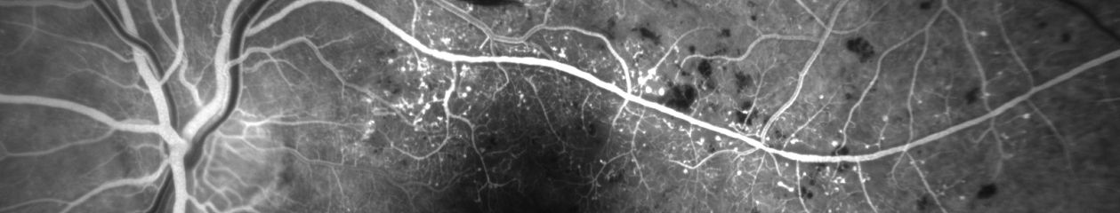
The most common uses of fluorescein angiography are in retinal or choroidal vascular diseases such as diabetic retinopathy, age-related macular degeneration, hypertensive retinopathy and vascular occlusions. Typically, these are clinical diagnoses and the angiogram is used to determine the extent of damage, to develop a treatment plan or to monitor the results of treatment. In diabetic retinopathy the angiogram is useful in identifying the extent of ischemia, the location of microaneurysms, the presence of neovascularization and the extent of macular edema. In age-related macular degeneration, angiography is useful in identifying the presence, location and characteristic features of choroidal neovascularization for possible treatment with laser photocoagulation, photodynamic therapy, or antiangiogenic medications.

Fluorescein angiography can be very useful in certain degenerative and inflammatory conditions. Some of these conditions exhibit characteristic fluorescence patterns, which support the diagnosis.

For example, Stargardts Disease exhibits a “silent choroid” and a central bulls-eye fluorescence pattern in the macula while acute posterior multifocal placoid pigment epitheliopathy (APMPPE) demonstrates a characteristic “block early, stain late” pattern.

Angiography also plays an important role in clinical research, advancing the understanding of retinal vascular disorders and potential treatment modalities. A number of multi-center clinical trials use fluorescein angiography to investigate new treatment options to combat retinal disease. As new therapeutic modalities are developed, fluorescein angiography will continue to play an important role in the management of common retinal conditions.
Dosage and Administration
 Fluorescein angiography is performed by injecting fluorescein sodium dye as a bolus into a peripheral vein. The normal adult dosage is 500mg, and is typically packaged in doses of 5 ml of 10% or 2 ml of 25%. For pediatric patients, the dose is adjusted to 35 mg per 10 pounds of body weight.9
Upon entering the circulation, approximately 80% of the dye molecules bind to plasma proteins, which significantly reduces fluorescence because the free electrons that form this chemical bond are subsequently unavailable for excitation.1 The remaining unbound or free fluorescein molecules fluoresce when excited with light of the appropriate wavelength. With a molecular weight of 376, fluorescein diffuses freely out of all capillaries except those of the central nervous system, including the retina.
The dye is metabolized by the kidneys and is eliminated through the urine within 24 to 36 hours of administration. During this period of metabolism and elimination, fluorescein has the potential to interfere with clinical laboratory tests that use fluorescence as a diagnostic marker.10, 11 To avoid any false readings, it may be prudent to schedule clinical lab tests either before the angiogram, or postpone testing for a day or two to allow sufficient elimination of the dye. Side effects of intravenous fluorescein include discoloration of the urine for 24 to 36 hours and a slight yellow skin discoloration that fades within a few hours. Nursing mothers should be cautioned that fluorescein is also excreted in human milk.12
Complications and Adverse Reactions
 Fluorescein sodium is well tolerated by most patients, but angiography is an invasive procedure with an associated risk of complication or adverse reaction. Use of fluorescein sodium may be contraindicated in patients with history of allergic hypersensitivity to fluorescein. Although generally considered safe for patients receiving dialysis, one manufacturer of fluorescein suggests using half the normal dose in dialyzed patients.13 There are no known risks or adverse reactions associated with pregnancy, but most practitioners avoid performing fluorescein angiography in pregnant women, especially in their first trimester.14-16
Historically, adverse reactions occur in 5-10% of patients and range from mild to severe. 17-23 Anecdotal evidence suggests a lower incidence of reaction in recent years and the first large study conducted in over a decade seems to confirm that, reporting a frequency of adverse reaction of just over 1% .24 Continued improvements in manufacturing processes and implementation of tighter pharmacopeial standards are credited with this reduction and may lead to lower rates of reaction in the future. 25
Transient nausea and occasional vomiting are the most common reactions and require no treatment. These mild reactions typically occur 30-60 seconds after injection and last for about one to two minutes. Fortunately, they seldom compromise the diagnostic quality of the angiogram. The incidence of nausea and vomiting seems to be related to the volume of dye and rate of injection. A relatively slow rate of injection often reduces or eliminates this type of reaction but can adversely affect image quality and alter arm-to-retina circulation times. Premedication with promethazine hydrochloride or proclorperazine may prevent or lessen the severity of nausea and vomiting in patients with a history of previous reactions to fluorescein, but is rarely needed and one study noted a higher frequency of these reactions in patients that had been premedicated. 21 Some patients report a strong taste sensation or hypersalivation following injection of fluorescein.
Moderate reactions occur less frequently, affecting less than 2% of patients that undergo angiography. Allergic reactions such as pruritus or urticaria can be treated with antihistamines, but any patient who experiences these symptoms should be observed carefully for the possible development of anaphylaxis. The advisability of performing angiograms in patients with a history of allergic reaction to fluorescein should be considered carefully, as allergic sensitization to the dye can increase with each subsequent use. Patients with previous history of mild allergic reaction to fluorescein can be pre-treated with an antihistamine, such as diphenhydramine, 30-40 minutes prior to any subsequent angiograms to limit allergic response, although this may not prevent serious reactions.26 Vasovagal attacks happen in some patients, 27 most likely due to anxiety about the procedure or their ocular condition. Usually the angiogram needs to be aborted or postponed, but some patients are able to tolerate the angiogram during the initial stages of a syncopal episode. However, the drop in blood pressure and heart rate can dramatically alter the angiographic results.28
More severe reactions are rare, but include laryngeal edema, bronchospasm, anaphylaxis, tonic-clonic seizure, myocardial infarction and cardiac arrest. 29-33 The overall risk of death from fluorescein angiography has been reported as 1 in 222,000.21 Although life-threatening reactions during angiography are rare, angiographic facilities and personnel should be properly equipped and prepared to manage serious reactions to the procedure. A resuscitative crash cart and appropriate agents to treat severe reactions should be readily available including epinephrine for intravenous or intramuscular use, soluble corticosteroids, aminophylline for intravenous use, oxygen, and airway instrumentation. It is generally recommended that a physician be present or available during angiographic procedures.
Extravasation of fluorescein dye during the injection can be a serious complication of angiography. With a pH of 8 to 9.8, fluorescein infiltration can be quite painful. If fluorescein dye extravasates, cold compresses should be placed on the affected area for 5 to 10 minutes, and the patient should be reassessed until edema, pain, and redness resolve. Serious complications are more likely to occur when large amounts of dye extravasate. Sloughing of the skin, localized necrosis, subcutaneous granuloma, and toxic neuritis have been reported following extravasation of fluorescein.34-36 To avoid these problems, continual observation of the injection site during the course of the injection and monitoring the patient for pain is recommended. Accidental arterial injections are rare, but can be quite painful. The dye remains concentrated and stains the effected extremity with little or no dye reaching the retinal vasculature. With proper technique, these complications of injection can usually be avoided.

In cases when venous access is severely compromised or the patient is known to be highly allergic to the dye, fluorescein can be administered orally.37, 38 Although oral fluorescein administration is typically well tolerated, severe adverse reactions can occur.39, 40 Due to the slow absorption rate, an early transit sequence is not possible. The resulting images are less than ideal, but have traditionally provided limited diagnostic information in conditions where late phase photographs are helpful, such as cystoid macular edema. The use of optical coherence tomography to detect the presence of edema or fluid within the retinal tissues has essentially eliminated this use of oral fluorescein.
For more on fluorescein angiography, visit:
Equipment & Technique
Interpretation
Step-by-Step
References:
- Wolfe DR. Fluorescein angiography basic science and engineering. Ophthalmology 93:1617-1620, 1986.
- Morris PF. Fluorescein sodium and indocyanine green: uses and side effects. In: Saine PJ, Tyler ME, eds. Ophthalmic Photography: Retinal Photography, Angiography and Electronic Imaging. 2nd ed. Boston, Butterworth-Heinemann, 2002:137-165.
- Jacobs J. Fluorescein sodium – what is it? J Ophthalmic Photography 14:62, 1992.
- Dole RB. Use of fluorescein in the study of underground waters. USGS Water Supply Paper 160:73-85, 1906.
- Myers B. Prediction of skin sloughs at the time of operation with the use of fluorescein dye. Surgery 51:158-162, 1962.
- McCaw JB, Myers B, Shanklin KD. The value of fluorescein in predicting the viability of arterialized flaps. Plast Reconstr Surg 60:710-719, 1977.
- Marfuggi R, Greenspan M. Intraoperative prediction of intestinal viability. Surg Gynae Obstet 152:33-35, 1981.
- Martonyi CL, Bahn CF, Meyer RF. Clinical Slit Lamp Biomicroscopy and Photo Slit Lamp Biomicrography. Ann Arbor, MI, Time One Ink, Ltd, 1985, pp 64-67.
- Physicians’ Desk Reference for Ophthalmic Medicines. Montvale, Thompson PDR;207, 2006.
- Palestine AG. Does intravenous fluorescein interfere with clinical laboratory testing?. J Ophthalmic Photography 13:27-28, 1991.
- Bloom JN, Herman DC, Elin RJ, et al. Intravenous fluorescein interference with clinical laboratory tests. Am J Ophthalmol 108:375-379, 1989.
- Mattern J, Mayer PR. Excretion of fluorescein into breast milk [letter]. Am J Ophthalmol 109:598, 1990.
- AK-FLUOR package insert, rev. 08/05. Akorn, Inc Buffalo Grove, IL.
- Halperin LS, Olk RJ, Soubrane G, et al. Safety of fluorescein angiography during pregnancy. Am J Ophthalmol 109:563-566, 1990.
- Greenberg F, Lewis RA. Safety of fluorescein angiography during pregnancy [letter]. Am J Ophthalmol 110:323-325, 1991.
- Berkow JW, Flower RW, Orth DH, Kelley JS. Fluorescein and Indocyanine Green Angiography, 2nd ed. San Francisco: American Academy of Ophthalmology, 1997 p 7.
- Chazan BI, Balodimos MC, Koncz L. Untoward effects of fluorescein retinal angiography in diabetic patients. Ann Ophthalmol 3:42, 1971.
- Pacirariu RI: Low incidence of side effects following intravenous fluorescein angiography. Ann Ophthalmol 14:32–36, 1982.
- Butner RW, McPherson AR: Adverse reactions in intravenous fluorescein angiography. Ann Ophthalmol 15:1084–1086, 1983.
- Marcus DF, Bovino JA, Williams D. Adverse reactions during intravenous fluorescein angiography. Arch Ophthalmol 102:825, 1984.
- Yannuzzi LA, Rohrer KT, Tindel LJ, et al. Fluorescein angiography complication survey. Ophthalmology 93:611-617, 1986.
- Karhunen U, Raitta C, Kala R. Adverse reactions to fluorescein angiography. Acta Ophthalmol 64:282-286, 1986.
- Kwiterovitch KA, Maguire MG, Murphy RP, et al. Frequency of adverse systemic reactions occurring after fluorescein angiography – results of a prospective study. Ophthalmology 98:1139-1142, 1991.
- Kwan AS, Barry CJ, McAllister IL, et al. Fluorescein angiography and adverse drug reactions revisited: the Lions Eye experience. Clin Experiment Ophthalmol 34:33-38, 2006.
- Eutick M. Sodium fluorescein – colourful past, bright future. J Ophthalmic Photography 28:66-70, 2006.
- Ellis PP, Schoenberger M, Rendi MA. Antihistamines as prophylaxis against side reactions to intravenous fluorescein. Trans Am Ophthalmol Soc 78:190–205, 1980.
- Buchanan RT, Levine NS. Blood pressure drop as a result of fluorescein injection. Plast Reconstr Surg 70:363-368, 1982.
- Merin LM, Lam BL. Case report: fluorescein angiogram during vasovagal syncope. J Ophthalmic Photography 16:94-95, 1994.
- Gombos GM, Lieberman RM. Seizures associated with fluorescein angiography. Ann Ophthalmol 21:89-90, 1989.
- Kelly SP, Mcdermott NJ, Saunders DC, et al. Convulsion following fluorescein angiography. Br J Ophthalmol 73:655-656, 1989.
- Hess JB, Pacirariu RI. Acute pulmonary edema following intravenous fluorescein angiography. Am J Ophthalmol 82:567-570, 1976.
- Deglin SM, Deglin EA, Chung EK. Acute myocardial infarction following fluorescein angiography. Heart Lung6:505-509, 1977.
- Acasco FJ, Tiestos MT, Navales J, et al. Fatal acute myocardial infarction after intravenous fluorescein angiography. Retina 13:238-239, 1993.
- Schatz H. Sloughing of skin following fluorescein extravasation.AnnOphthalmol 10:625, 1978.
- Elman MJ, Fine SL, Sorenson J, et al. Skin necrosis following fluorescein extravasation. A survey of the Macula Society. Retina 7:89-93, 1987.
- Lipson BK, Yannuzzi LA. Complications of fluorescein injections. Int Ophthalmol Clin 29:200-205, 1989.
- Kelley JS, Kincaid M. Retinal fluorography using oral fluorescein. Arch Ophthalmol 97:2331-2332, 1979.
- Hara T, Inami M, Hara T. Efficacy and safety of fluorescein angiography with orally administered sodium fluorescein. Am J Ophthalmol 126:560-564, 1998.
- Kinsella FP, Mooney, DJ. Anaphylaxis following oral fluorescein angiography. Am J Ophthalmol 106:745-746, 1988.
- Gomez-Ulla F, Gutierrez C, Seonae I. Severe anaphylactic reaction to orally administered fluorescein. Am J Ophthalmol 112:94, 1991.