
In 1959, two medical students, Harold Novotny and David Alvis began working under John Hickam, Chairman of Medicine at Indiana University on a research project funded (in part) by the United States Air Force. Dr. Hickam was interested in the retinal vascular bed as a visible segment of the cerebral circulation. He was searching for a photographic technique to estimate blood oxygen concentrations in retinal vessels.1
The students began by learning to perform fundus photography and they noted what appeared to be fluorescence from the crystalline lens. This led them to question whether it would be possible to photograph fluorescence in the retinal vessels using a fluorescent dye such as fluorescein. They presented the idea to Dr. Hickam, who gave his approval along with the suggestion that they would need to determine the excitation and emission wavelengths of fluorescein. A blood sample was mixed with fluorescein and sent to James Hartigan at the Eli Lilly Pharmaceutical lab for measurement with a spectrofluorometer. They found the peak excitation of fluorescein to be around 480nm and peak emission to be 520nm.

They then plotted the spectral transmission curves of numerous filters looking for a combination that would be effective for angiography and chose a Kodak Wratten 47 blue filter for the exciter and a Wratten 58 green for the barrier. They added a copper sulfate water bath solution as an adjunct to the barrier filter and felt that it was not essential, but resulted in sharper images. Ironically, the blue color of copper sulfate probably made their filter system less efficient and increased the crossover in the transmission curves of the broadband absorption filters.
Using Ansco Super Hypan 500 speed film (pushed to 2400), they first tried the technique on rabbits and then reportedly flipped a coin to see which one of them would be the first “patient”. Alvis lost, so he was the subject of the first successful fluorescein angiogram in a human!

Following their initial results, they refined the technique on diabetic and hypertensive patients in the ophthalmology clinic as part of their research project. Neither Novotny, nor Alvis had ever written for publication before, but completed a manuscript hoping to get published in the ophthalmic literature. A brief abstract from their presentation to the Midwest ARVO meeting (April 23-24, 1960) appeared in the American Journal of Ophthalmology (AJO) in 19602 and a technical report submitted to the USAF School of Aviation Medicine (the sponsoring agency) was published in September 19603, but a full manuscript submitted to the AJO was rejected as unoriginal work! The reviewers cited work previously published by Milton Flocks and coworkers who had used a 16mm movie camera and a carbon arc light source to obtain fluorescein angiograms in cats, but had been unsuccessful in humans due to insufficient light.
After being rejected by the ophthalmic literature, Dr. Hickham forwarded the manuscript to a colleague who was editor of the journal Circulation. The landmark paper, “A Method of Photographing Fluorescence in Circulating Blood of the Human Retina.” was published in Circulation in 1961.4

Novotny and Alvis went on to continue their medical education. Alvis began a residency in ophthalmology at Wayne State University, where he made several requests for support to continue his work in angiography. His requests were denied and he did not pursue angiography any further.
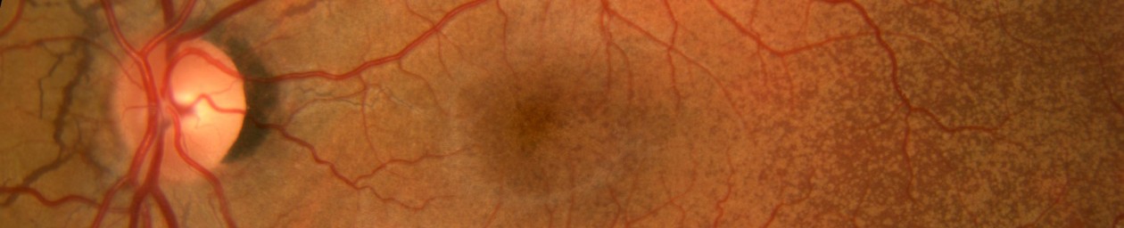
Following publication of their report, several investigators studied various clinical applications of angiography, quickly establishing the intrinsic value of the technique. Further technical innovations such as improved filter combinations, fast recycling flash units, and digital imaging techniques were gradually implemented, but the basic principles and technique originally described by Novotny and Alvis remain the same today.
Harold Nonotny went on to pursue a career in psychiatry. Alvis completed his training and practiced as a general ophthalmologist. He never utilized fluorescein angiography in his practice. Some years later however, Alvis was invited to speak at the International Symposium on Fluorescein Angiography where was recognized for his pioneering role in fluorescein angiography and received an apology from the journal editor who had originally rejected the manuscript. Finally feeling some vindication for his efforts, Alvis wrote, “I had an idea how Orville and Wilbur Wright must have felt when they finally received support for their heavier-than-air machine which was thought to be unworkable.” 5

Novotny also humbly took a little credit for his role in pioneering angiography. In 2003, I was doing data entry on a survey conducted by the Ophthalmic Photographers’ Society and came across this response from Harold Novotny!

As Honorary Life Members, Novotny and Alvis were both on the OPS mailing list. Dr. Novotny marked his credentials as MD and simply replied, “Created Fluorescein Angiography”!
This story demonstrates that research doesn’t necessarily follow a straight line to scientific discovery. Novotny and Alvis were looking for a method to estimate blood gas concentrations for the USAF and ended up developing techniques for fluorescein angiography. Great discoveries can sometimes start from very modest beginnings.
References:
- Alvis DL. Twenty-fifth anniversary of fluorescein angiography. Letter to the Editor. Arch Ophthalmol 1985; 103:1269
- Novotny HR, Alvis DL. A method of photographing fluorescence in circulating blood of the human retina. Abstract from Midwest ARVO Meeting. Am J Ophthalmol 1960 50:176.
- Tredici TJ. History of fluorescein angiography corrected. Letter to the Editor. Arch Ophthalmol 1986; 104:21.
- Flocks M, Miller J, Chao P. Retinal circulation time with the aid of fundus cinematography. Am J Ophthalmol 1959; 48:3-6.
- Novotny HR, Alvis DL. A method of photographing fluorescence in circulating blood of the human retina. Circulation 1961; 24:82. http://circ.ahajournals.org/content/24/1/82.full.pdf+html
- Alvis DL, Julian KG. The story surrounding fluorescein angiography. J Ophthal Photography 1982; 5:6-8