Tag Archives: fundus photography

Solving the Puzzle of History


Our profession enjoys a long and rich history, one that has seen dramatic advancement in technology and techniques that aid in the documentation and diagnosis of eye disease. My personal interest in the history of ophthalmic photography stems from participation in multiple history symposia sponsored by the  American Academy of Ophthalmology’s Museum Committee. In 2011, I was quite honored to be invited to co-chair the symposium Imaging and the Eye, but I think I drew the short straw when we were assigning lecture topics. I was given the task of covering the origins of photography and ophthalmic photography – in a ten minute presentation!

It seemed like a daunting task to somehow cover all that history in such a short talk. But when I started to research the topic, a common theme began to emerge when I came across rivalries, controversies, mistakes, and inconsistencies in the historical accounts related to several important discoveries. Rather than try to force every important milestone or event into a timeline format, I decided to concentrate my lecture on a few important controversies and rivalries. This approach worked well for the symposium, but I ended up with far more material than I could hope to cover in several lectures. And I had barely scratched the surface.

The Pencil of Nature is the first collection of photograph works published in book form by Fox Talbot in 1844.  The photographs were printed separately and then mounted or “tipped in”. Talbot claimed that his photographic process preceded that of Daguerre’s in 1839. A digitized version of this book is available through Project Gutenberg.

During the research process, I found that reconstructing history is somewhat like completing a puzzle. Professional historians traditionally have had access to original source documents to support their historical research. Thanks to digital technology, many of these obscure resources are now publicly available through advanced search engines and extensive online collections of scanned historical journals and documents. New pieces of the historical puzzle often become apparent when you can access these primary documents. The accounts in this series benefit from the availability of newly digitized documents, many of which were originally published over 100 years ago.  The Internet Archives, Project Gutenberg, and Google Books provide access to digitized, publicly accessible books, periodicals, and journals that are now in the public domain by virtue of their age and expiration of copyright.

Advertisements in The Philadelphia Photographer from June, 1886. Jackman and Webster’s landmark article on the first successful fundus photograph is included in this issue of the photographic periodical. It can be accessed and downloaded from the Internet Archives.

Even with access to these amazing resources, there are still some missing pieces of the puzzle. The available literature sometimes contains conflicting information or apparent mistakes between different historical accounts. Some publications have also proven to be difficult to locate, either online or in print. These hard to find references were often published in the decades just prior to routine digital publication (1960’s & 70’s) and may not yet be eligible for inclusion in public domain collections.

Covers from vintage journals accessed through Google Books. The 1894 Transactions of the Ophthalmological Society of the United Kingdom includes a paper on fundus photography by one of the early pioneers, Lucien Howe. The American Journal of Ophthalmology from 1899 includes a description of Thorner’s reflex-free ophthalmoscope. Thorner and others soon adopted the same optical design to build improved fundus cameras.

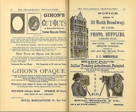
In piecing this puzzle together, I found that it pays to read all referenced documents that other historians have cited rather than rely on a citation of a “fact” actually being accurate. Mistakes are sometimes made and then blindly repeated or misinterpreted in other accounts. For example, a non-existent reference title was accidentally published in multiple historical reviews. Listed as “Barr E.: Drs. Jackman & Webster, Philadelphia Photographer June 5, 1886”, it combined fragments of two separate references and was most likely an author’s note to search for them both. 1,2

After searching the online archives, I was able to confirm that the combined title doesn’t exist, yet multiple authors include it in their reference list.3,4  The authors may have also been confused because of a typographical error in multiple references. Elmer Barr was listed as author of an 1887 paper in the American Journal of Ophthalmology, as well as another article in the Scientific American Supplement from 1888.1,5  Both of these articles describe the successful capture of a human fundus photo with more recognizable features than previous investigators. The author’s real name was Elmer Starr, but the typographical error was repeated several times causing an early pioneer in fundus photography to fade into obscurity and lose his rightful place in history. Being able to detect these mistakes and correct the historical record of our profession has been fascinating.

Die Photographie des Augenhintergrundes by Friedrich Dimmer is an atlas of fundus photographs, published in 1907. It contains several amazing reflex-free photographs taken with a one-of-a-kind camera of Dimmer’s own design. This book represents a major milestone in ophthalmic photography. It was digitized by The Internet Archives in 2011 with funding from Open Knowledge Commons and Harvard Medical School.

In piecing these puzzles together, what stood out the most were the often bitter rivalries that seemed to overshadow many of the most important discoveries. Photography was born in the Victorian Era, a time of great discovery, invention, and advancement in science and medicine. The Victorian Era roughly coincided with the Belle Epoch in Continental Europe and the Gilded Age in the United States. It was during this period that Darwin, Babbage, Pasteur, Maxwell, Morse, Helmholtz, and many others made important advancements in science, medicine, and technology. As you will see in future installments of this series, it was also a time of fierce competition, rivalry, and controversy. The brilliant minds of the day often had egos to match their great intellect. The race to be listed as the “first” to discover a scientific breakthrough could become an obsession. Eponyms were popular, and just about every important new discovery was named for the person that first described it.

A classic example of this competition and controversy occurred in the feud over the discovery of anesthesia in the 1840’s when American dentist Horace Wells and his former apprentice William Morton both claimed to be the first to discover the use of inhaled anesthesia. Wells had successfully used anesthesia on several occasions, but was discredited after a famously failed public demonstration. Humiliated after this one failure, he became deeply depressed, began abusing chloroform, and eventually committed suicide. Morton didn’t fare much better. He remained obsessed with recognition throughout his life. He tried to patent ether under a different name, and eventually died penniless. The American Dental Association honored Wells posthumously in 1864 as the discoverer of modern anesthesia, and the American Medical Association recognized his achievement in 1870. Morton was similarly recognized later in life and again posthumously. Both were instrumental in this major medical advancement, but their egos prevented them from sharing in recognition of their achievement.

The next few episodes in this historical series explore similar relationships, rivalries, feuds, and debate surrounding several important milestones in the evolution of ophthalmic imaging. Fortunately the ending of each of these stories is slightly less morbid than the anesthesia saga:

The Priority Debate looks at the frantic race for recognition as the inventor of photography in 1839.

Stereo Photography examines the nineteenth century development of the stereoscope and competing theories on stereo vision that resulted in a bitter feud between Wheatstone and Brewster.

The First Human Fundus Photograph will explore several controversies and professional rivalries in the early days of fundus photography, including how Elmer Starr lost his place in history, as well as another rivalry that led to accusations of falsifying photographic results.

From there we will continue to explore the evolution of ophthalmic imaging by taking a look back at important individuals and events that shaped our field – and hopefully fill in a few more pieces of the historical puzzle that represents the legacy of our profession.

References:

  1. Barr E. On photographing the interior of the human eyeball. Amer J Ophth 1887; 4:181-183
  2. Jackman WT, Webster JD. On photographing the retina of the living eye. Philadelphia Photographer 1886;23:340-341
  3. Van Cader TC. History of ophthalmic photography. J Ophthalmic Photography 1978; 1:7-9
  4. Wong D. Textbook of Ophthalmic Photography. Inter-Optics Publications, New York, 1982
  5. Barr E. Photography of the human eye. Scientific American Supplement 1888; 650:10388

Book Review

Optical Coherence Tomography and OCT Angiography: Clinical Reference And Case Studies

Darrin A. Landry, CRA, OCT-C
Amir H. Kashani, MD, PhD

Bryson Taylor Publishing, 2016
ISBN–‐13:978–‐1523976867
ISBN–‐10:1523976861
www.BrysonTaylorPublishing.com

In the early days of retinal angiography, photographers often worked very closely with ophthalmologists, learning together as they explored the diagnostic uses of fluorescein angiography and unraveled the complexities of interpreting the fascinating images they were capturing. This spirit of scholarly collaboration between imager and physician continues today in a new book: Optical Coherence Tomography and OCT Angiography, Clinical Reference And Case Studies by Darrin Landry and Amir Kashani. These authors are both well respected in their respective fields as educators and authors. Together they have created a timely textbook that will appeal to members of both professions.

Before receiving an advance copy of this book for review, I anticipated that the content would focus almost exclusively on OCT angiography.  I was pleasantly surprised to find that although the book features OCT-A prominently, it is much more than a text on this new state-of-the-art technology. It appropriately places OCT-A in the context of multiple imaging modalities to assist in diagnosis of a variety of retinal conditions.

The authors have produced a book that is part tutorial, part clinical atlas, and a collection of over forty cases that “puts it all together” using multiple imaging modalities with clinical descriptions. The book is divided into three sections:

Section 1. OCT and OCT Angiography

The introductory section will be particularly useful to imagers as it includes a basic overview of OCT and OCT-A technology, followed by a discussion on pattern recognition, normal anatomy and layers of the retina, how to move the scan pattern, recognizing artifacts, EDI/FDI and a basic primer on OCT-A. The OCT-A primer explains how this technology scans through the z-axis and detects motion to identify the retinal vasculature including the deep retinal plexus.  It includes a discussion of artifacts specific to OCT-A . This section will be especially helpful to those new to OCT and OCT-A, and anyone preparing for certification as an OCT-C.

Section 2. Atlas of Images and Disease Pathology

This section is an atlas of retinal OCT findings organized in anatomical order from the vitreous to the choroid. For each condition, the text includes a brief discussion of the disease process, clinical findings, and appearance in multiple modalities. For each condition, there are multiple image examples providing a full spectrum of potential findings for that disease. For instance, there are over twenty different examples of epiretinal membrane. Novice imagers will find this variety especially helpful in learning to recognize different manifestations of a single condition. In addition to common retinal findings the book also includes good examples of less recognized conditions such as outer retinal tubulation (ORT) and reticular pseudodrusen. As expected, retinal vascular diseases include OCT-A examples along with SD-OCT and other imaging modalities including fluorescein and ICG angiography. Experienced imagers will recognize many of these conditions, but the addition of OCT-A will give them another viewpoint and expand their understanding of each disease.

Section 3. Case Studies

The final section of the book is a series of over forty cases where the authors combine a brief medical summary with appropriate imaging modalities for clinical correlation. This format fits well with the current trend of “case-based-learning” in medical education. In many of these cases, OCT-A dovetails nicely with other imaging modalities to increase our understanding of a disease process or help confirm a diagnosis. This quote from the book’s Preface describes the format well “These images are presented in the context of additional imaging modalities to aide the reader in making useful correlations.”

In conclusion, this timely book is well organized and thorough, without becoming unwieldy. It is easy to navigate between sections if you want a quick reference on OCT anatomy or to look for examples of specific retinal conditions and how they may appear on OCT, OCTA and other imaging modalities. With over a thousand images and forty cases, to say that this book is generously illustrated would be an understatement. It is an impressive collaboration between an ophthalmic imager and a retinal specialist that should appeal to a wide audience that would include ophthalmic imagers, retinal technicians, residents in training, and clinicians wanting a reference for clinical correlation between modalities.

From a personal standpoint, I think it’s great to have an ophthalmic imager making a significant contribution to the ophthalmic literature. Darrin’s collaboration with Dr Kashani serves as a model for what imagers can accomplish when we collaborate with physicians on a professional level.  The spirit of collaboration between professions is echoed several times in the book including this statement from the Introduction, “Constant and close communication between the physician and imager is very essential.”

Reviews like this often end with a cliché that suggests that everyone in the profession should “add this book to your collection” or “keep a copy on your bookshelf”. I’ve tried to avoid those clichés, but honestly, I am happy to have this book in my collection and plan to keep it handy in clinic for reference, especially as we integrate OCT-A into our own diagnostic armamentarium.


To Blink, or not to Blink?

It seems almost too obvious to mention, but just like you can’t see through a window when the window shade is pulled down, you cannot view or image the interior of the eye through closed eyelids.

close-open1-640

Obviously we need fully retracted upper and lower lids to get the best view of the fundus with our fundus camera, SLO, or OCT. Because these are noncontact imaging techniques, image quality is also dependent on a regular ocular surface and clear ocular media. An intact tear film is an important optical component of the ocular media. Simply put, to get the best images we need to strike a balance between fully retracted lids and frequent blinking to maintain the tear film.

miller pre-post-640
Top: Shadow from partially retracted upper lid appears at the bottom of the fundus image and degrades the OCT signal. Bottom: fully retracted lid improves the illumination of fundus image and improves signal strength in the OCT.
mccalister pre-post 640
Lashes partially obscure the retinal view in the top image. A fully retracted lid improves the view.

Many patients are nervous about their visual symptoms and what diagnosis the imaging procedure might detect. They often try hard not to blink during the session thinking it will help you get the best images. But their tear film will break up during this time and the view will become compromised until they blink again. And they often apologize for blinking!

To compound this dilemma, these imaging tests are often performed after a patient has undergone an extensive screening workup that includes IOP measurement, and application of topical anesthetic and dilating solutions. Patients may also undergo gonioscopy or macular contact lens examination prior to imaging. A disrupted tear film is an unintended side effect of these procedures and can adversely affect imaging quality.

pre-post blink 640
Top: irregular ocular surface causes degradation of both the fundus image and OCT as the tear film breaks up from lack of normal blinking. Bottom: after a few blinks, the view improves dramatically. Artificial tears would similarly improve the view.

It may seem counter-intuitive, but encouraging patients to blink frequently during imaging sessions can improve cooperation and image quality in fundus photography and OCT imaging. In our clinic, patients are often surprised that we encourage them to blink, having had procedures done in other clinics where they were sternly cautioned against blinking. In my experience as a consultant and workshop instructor, I have often heard OCT operators repeat the words “Don’t blink!” while performing a raster scan pattern that may take several seconds to capture.

cirrus blink2-640
Artifact from blinking during a volume scan. Timing the patient’s blinking pattern can avoid this type of artifact.

They know that a blink will result in an artifact in the volume map, but fail to recognize the need for frequent blinking. I don’t really blame the operator. Often that’s how they were taught to perform the scan during a workshop or training session by the manufacturer’s trainer:

“Don’t blink! Don’t blink! Don’t blink! Don’t blink! Don’t blink! Don’t blink! Don’t blink!….”

No wonder the patients are afraid to blink! Frequent blinking not only refreshes the tear film, it makes the patient feel more comfortable and ultimately more cooperative. You’ll soon learn to recognize a patient’s blinking rhythm and you can time your image capture just as their upper lid is retracting after a blink. Gently encourage the patient by saying, “hold your gaze for just a moment” when you need just a second or two longer to capture a good image. When frequent blinking doesn’t work, application of artificial tears can also make a difference in patients with dry eyes or compromised tear film.

blink clark 4-up-640
Partial versus fully retracted upper eyelid. Image quality is compromised by the lid and lashes in the left images. Gently retracting the upper lid immediately after the patient blinked improves image quality.

During fundus photography, the flash of the camera will cause an involuntary blink that helps refresh the tear film. If the lid or eyelashes obscure the view, gentle retraction of lids with a finger or q-tip may help. You don’t need to forcefully tug on the lid, just retract it a couple of millimeters to get any lashes out of the way and reveal the entire pupil. Patients are often still able to blink with this mild retraction of the upper lid.

So encourage your patients to blink regularly and learn to capture the best images in between the blinks. If it weren’t for all the blinks, anyone could do this job!

First Look: Eidon Retinal Scanner

I recently had the chance to get a hands-on look at the Eidon confocal retinal scanner.  The Eidon is a hybrid device combining features of a non-mydriatic fundus camera with confocal scanning technology. It is manufactured in Italy by Centervue SpA. Centervue describes this instrument as the first true color confocal scanner on the market. It is different than a confocal scanning laser ophthalmoscope in that it uses a broad spectrum white light LED (440-650 nm) rather than monochromatic lasers.  A second light source provides near infrared (IR) imaging at 825-870 nm. The advantage to confocal imaging is that it suppresses out-of-focus light from reaching the image sensor. This minimizes the effect of cataracts or other media opacities, resulting in sharp, high contrast images. The confocal design also allows it to image through a smaller pupil than a typical non-mydriatic camera.

eidon1The footprint of the Eidon is fairly compact, but the instrument is taller than most fundus cameras. The device is operated via touch screen tablet and has both automatic and manual controls.  The Eidon has a fixed 60 field of view, but is capable of capturing several fields and creating montage images. It features a 14 megapixel sensor to capture color, red free, and infrared images. The red free photos are extracted from the color image rather than through a separate exposure with a blue-green light source.

The capture software is incredibly simple to use. It is about as automatic as a device can get. Using the touch screen tablet, you enter the patient demographics and program it for the desired fields of one or both eyes and push the start button. The device does the rest automatically, even telling the patient to open their eyes prior to each flash capture.

Eidon2

The internal fixation light will step through the various fields and capture each one automatically. Auto-alignment is accomplished by identifying the center of the patient’s pupil with IR.  It will then focus automatically with a range of -12D to +15D. Once focused in IR, the camera will slightly readjust focus just prior to color capture to account for the difference in wavelengths between color and IR. The autofocus works very well, but eye movement during capture can contribute a slight blur to the image.

Minimum pupil size is 2.5mm. It does capture good images at this pupil size in the posterior pole view but like any other non-myd device, it works a little better if patients are pharmacologically dilated. This is especially helpful when imaging peripheral fields or you plan to do a montage. I have found this to be true with all non-myd color fundus cameras. I would like to see separate exposure settings to reduce the gain and noise for eyes with widely (pharmacologically) dilated pupils.

composite 4-640
Left to right: cropped images from a non-mydriatic camera, Optos composite red/green, and Eidon. Photos of the same pseudophakic eye were taken on different dates.

The resulting images appear different than what we see with either a digital fundus camera or a cSLO. Centervue refers to the broad spectrum imaging as “True Color” to distinguish Eidon images from SLO composite laser color images from Spectralis or Optos.

todd darker 640

The Eidon attempts to address some of the limitations of digital fundus cameras that are poorly calibrated for color balance, gamma, and exposure. In doing so, it seems to sacrifice some color fidelity and a true appearance of the optic nerve. The red channel is desaturated to avoid loss of detail from oversaturation, but many Eidon images appear slightly green and might benefit from a little more red or magenta bias to the color balance.

noise1

Although the pixel count of the Eidon sensor is quite high, the color images seem a little over-processed and a bit noisy when zoomed in, probably from  the increased contrast as well as the high gain settings that allow it to capture through very small pupils.

cropped nerve
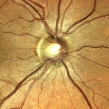
One of the features touted by the manufacturer is that it prevents “optic disc bleaching” seen with some fundus cameras. It does hold detail in optic disc photos, but the flip side to this is that the rim of the nerve can appear abnormally dark or gray, making it difficult to document pallor. Disc bleaching shouldn’t be  a problem in fundus cameras that are calibrated for proper contrast and exposure.

disc compare2-640
Left: Traditional color fundus image with a well balanced 11 MP color sensor. Right: Same eye taken with Eidon.

I also played with the digital joystick and manual mode changing the level of focus to see if the instrument exhibited the confocal tonal shift seen with the Spectralis. In playing with manual mode to alter focus or exposure, it became clear that the instrument works best in full-auto mode.

ICSC3-640
We did not see the confocal tonal shift in either color or IR images when looking at elevated lesions or manually changing the focus. Left: Spectralis IR (820 nm) image of serous detachment exhibiting tonal shift from elevation. Right: Eidon IR (825-870 nm) does not demonstrate the same effect even though it is also a confocal device.

The Eidon review software is functional, but could be a little more streamlined. It would be nice to scroll though successive images, rather than having to go back and forth to the proof sheet to open each frame individually. The montage software works quite well.

montage darker 640

The bottom line is that the Eidon is a very interesting hybrid device that combines features of confocal scanning with full color capture in a package that is incredibly simple to use. It would be a great screening tool or replacement for a fundus camera in primary eye care settings and would require minimal staff expertise or training.

Thanks to Todd Hostetter, CRA, COMT for bringing the device to the clinic for a demo, and to Jim Strong, CRA, OCT-C for help taking some of the images.

Disclaimer: I have no financial or proprietary interest in this device.

Mystery Diagnosis: Chairman’s Challenge

One of the cool things about being an ophthalmic imager is the teamwork that can develop with the ophthalmologists you work for. They rely heavily on the images you provide to help diagnose various eye conditions. I’ve been fortunate to work with physicians that rely not only on my imaging expertise, but also my experience in recognizing clinical features of unusual conditions. Recently, one of our medical retina specialists (my boss) challenged my diagnostic skills with an unusual case.

challenge stickyThe photo request came through as a post-it note that simply listed the patient’s name and the words: “Fundus photos & OCT”. There was no diagnosis listed or specific instructions given. I called the patient in to the imaging suite and it was a teenage girl accompanied by her mother. I asked if the doctor had given them any paperwork for me and the patient’s mother said, “No. Doctor Q said he wanted to see if you could guess the diagnosis”. Dr. Q and I have worked closely together for twenty years and I can often anticipate exactly what type of images he needs without much direction. But this was unusual. The lack of information was clearly deliberate on his part, which got me thinking:

Is he testing me?

Or does he need my help in making the dx? Yeah that’s it.

Wait… He’s Chairman of the Department, a sub-specialist in medical retina, has written a textbook/atlas of retinal diseases….

He’s clearly testing me, but it must be a rare condition. Hmmm….

Aha! He probably wants images for his new book.

These thoughts are swirling through my mind as I start with an OCT of the right eye. The IR fundus image shows a large oval area that is dark from elevation. It’s suggestive of a serous retinal detachment, but the OCT shows no fluid. The retinal pigment epithelium is pushed forward suggesting a choroidal lesion.

OsteomaA

I start going through a differential diagnosis process in my mind as I continue to capture images:

Nope, not a serous detachment. There’s increased choroidal reflectivity and thickening which suggests a choroidal tumor or nevus of some type. Could it be a melanoma or an osteoma?

Osteomas are pretty rare. I’ve seen one case in the past 30 years. Probably not…

I move to the fellow eye and the OCT shows significant pigmentary changes, subretinal fluid, and what looks like a choroidal neovascular membrane.

OsteomaB

Hmmm…. The findings are more dramatic in the left eye. Looks like maybe an exudative process in a young patient … could it be Coat’s Disease?

I move the patient to the fundus camera and peer through the eyepiece.

osteomaR

Yikes! I wasn’t expecting that. Maybe I was right to think Coat’s.

Coat’s disease is an idiopathic developmental retinal vascular abnormality in children.

coats1

Characteristic findings include telangiectatic vessels with aneurysmal dilation and exudative detachments.

But Coat’s doesn’t quite fit in the right eye. Maybe Vogt Koyanagi Harada disease (VKH)?

VKH1

Features of VKH include: choroidal thickening, hyperemic optic disc, multi-lobed serous detachments and de-pigmentation and clumping of the RPE late in disease.

Although some of the features fit, the characteristic serous detachments of VKH aren’t present. Both Coat’s and VKH are unusual, but not rare. At least not rare enough for Dr. Q to challenge me like this.

I shifted the camera to the left eye to take photos. Once again I was surprised at the clinical appearance.

osteomaL

At that point I must have smiled a little because the patient’s mom asked if I knew the condition. I said yes. Although it looked somewhat like Coat’s, I was almost certain it was a case of bilateral choriodal osteomas. A moment later, Dr. Q came into the imaging suite and confirmed the diagnosis as choroidal osteoma. Sure enough, he wanted good photos for his next book!

osteoma5

Choriodal osteomas are benign ossifying tumors of the choroid composed of mature bone elements. They often demonstrate a thin plaque-like yellow-tan lesion in the macula with sharp, scalloped borders. They are usually unilateral, but can be bilateral. Symptoms include metamorphopsia, scotoma and blurred vision. Vision loss may be due to direct tumor involvement or secondary to choroidal neovascularization with subretinal fluid, lipid, or hemorrhage.

Now that the mystery was over and I had passed the test, Dr Q. handed me the patient’s chart as I finished up the photo session. Her clinical findings were listed:

  • 15 y.o. female
  • VA: 20/25 OD 6/200 OS
  • IOP: 15/10
  • Pupils: Left APD (afferent pupillary defect)
  • SLE: WNL OU
  • DFE:
    • OD: Orange placoid elevated lesion
    • OS: Yellow-orange lesion with well-defined borders. Pigmented CNVM w/ subretinal hemorrhage
  • B-scan ultrasonography demonstrated classic highly reflective plaque-like structures with an acoustically empty region behind the tumors.

osteoma8

This case prompted me to look up the previous case I had encountered nearly twenty years ago. It was before the advent of OCT, but the osteoma was well documented with multi-spectral monochromatic imaging, color fundus photography, fluorescein angiography and B-scan ultrasonography. Dr. Q included that case in his retinal atlas and I had used it lectures on multi-modality imaging. The clinical appearance was quite striking and unforgettable.

osteoma12

It’s great when physicians challenge their staff to take an active role in a team approach to eye care. I’m fortunate to work with physicians like Dr. Q, who challenge me to not only capture high quality images, but to recognize the clinical features of routine and rare cases. Luckily, I passed this test!