Tag Archives: SLO

From Blogger to Scholar?

I always look forward to receiving my copy of the the Journal of Ophthalmic Photography in the mail. Even after reading digital proofs as an editor or contributing author it’s great to see the final product in print. There’s just something tangible and authentic about turning the pages of a high quality publication printed on good stock.

I was especially looking forward to the Spring 2016 issue of the JOP. It contains an article I contributed on The Confocal Tonal Shift. What’s unique about this article is that evolved from a blog post here on eye-pix into a scholarly article (of sorts).

The blog was based on an observational series of photographs that documents the tonal changes that occur when focusing the Heidelberg Spectralis, a confocal scanning laser ophthalmoscope (cSLO). I put the blog together to further my own understanding of just what I was seeing with the cSLO, and share my observations with fellow imagers. When asked to convert the blog into a piece for the JOP, I initially felt it was too informal, opinionated, and lacking strong references to be included in the professional literature.

web image2-640 crop

But the kernel of a good idea was definitely there. So I worked backwards and did a literature search after the fact. Then I reworked the article and was able to place my observations into the context of other clinical findings described in the ophthalmic literature. Probably not the ideal way to develop an idea into an article, but in this case it worked.

A mentor of mine has always encouraged me to get as much mileage as possible out of a project. The Confocal Tonal Shift is a good example of that. It’s gone from a blog post to a formal article in a well-respected professional journal. I’ve also adapted it into an expanded lecture topic (The Quirks of Confocal Imaging) that  I presented at a recent educational program. It received good feedback and will work it’s way into my lecture rotation this year.

The full journal article can be viewed here.

Bennett TJ. The confocal tonal shift.
Journal of Ophthalmic Photography, 38(1):17-22, Spring, 2016

The Confocal Tonal Shift

The Heidelberg Spectralis confocal scanning laser ophthalmoscope (cSLO) is a commonly used diagnostic imaging device that uses monochromatic laser illumination to image the eye. It can be used for several retinal imaging modalities including infrared reflectance (IR), fluorescein angiography, ICG angiography and fundus autofluorescence (FAF). The confocal capability of the cSLO allows it to capture high-contrast, finely detailed images.

But what does confocal actually mean and how does it work? The word confocal simply means “having the same focus”. In this case it refers to the confocal pinhole or aperture that is optically located at the same plane of focus as the subject. The cSLO utilizes a focused laser to scan the subject point-by-point and then captures the reflected light after it passes through a confocal pinhole. The pinhole suppresses out-of-focus light from reaching the image detector resulting in very sharp images. The confocal pinhole is especially effective at eliminating unwanted scatter from cataracts or corneal opacities since these structures fall far outside the plane of focus.

cataract
Left: Patient with a cataract obscuring the view of the retina and optic nerve through a fundus camera. Right: The confocal pinhole of the cSLO suppresses the scatter from the cataract improving the view of the fundus.

When imaging a patient, you can see the confocal effect as you adjust the focus to the plane of the retina where it is most light efficient. The image on screen will get brightest just as you come into sharpest focus. A secondary effect of the confocal aperture is how it effects the appearance of elevated or out of focus retinal structures.

papilledema1
The plane of focus effects reflectivity and appearance of retinal tissues based on depth due to the confocal aperture. All three images are at the same wavelength. Focus is on the elevated optic disc on the left image. Tonality changes as focus is shifted to the retinal surface.

Adjusting the focus knob of the Spectralis can have a dramatic effect on the tonal appearance of elevated structures such as papilledema or vitreous floaters as seen here in this video.

Note the optic nerve get progressively darker as focus is adjusted from the peak of the nerve to the surface of the surrounding retina, which starts to appear brighter. The opposite occurs in the second example. Vitreous floaters from asteroid hyalosis appear as dark shadows when focus is set on the optic nerve. As focus is shifted up into the vitreous, the floaters begin to brighten and the retina fades to  dark. The brightness/exposure has not been adjusted during this tonal shift.  The only change is the focus.

So what does this mean for us as diagnostic imagers? Because of the inherently shallow depth of focus of the cSLO, some ocular structures may appear dark simply because they are slightly out of focus. Elevated serous detachments or papilledema are examples of this phenomenon that I call the confocal tonal shift.

serous detachment 2
A case of central serous chorioretinopathy with a classic serous detachment that can be seen ophthalmoscopically. The cSLO image is dramatic in it’s appearance due to the confocal shift. The dark area is elevated and filled with clear fluid (not blood).

In some cases the confocal tonal shift can enhance the diagnostic information by clearly outlining the borders of an elevated area or lesion. The effect is most notable with the IR laser and in red free mode.

wavelength small
The confocal tonal shift in three different modalities. From left to right: IR, monochromatic blue, fundus autofluosescence. The effect seems to be most pronounced in IR mode.

The confocal tonal shift also has the potential to create tonal “artifacts” which can confound the appearance of findings like blood or hemorrhage that inherently appear dark.  Vitreous opacities will appear dark because they are usually out of focus and blocked by the confocal pinhole. But are they from blood or vitreous debris? It’s impossible to tell with the cSLO since they appear the same even though one is translucent and one is more opaque when viewed ophthalmoscopically or with a fundus camera.

vitreous floaters-640
Vitreous debris can appear very dark in cSLO images even though it is almost completely transparent. Without a frame of reference, it is impossible to know if these dark areas represent floaters or vitreous hemorrhage.

Similarly, it is difficult to distinguish between blood and elevation within retinal tissues in conditions such as macular degeneration, retinal vein occlusions and diabetic macular edema.

NPDR2-640
Patient with bilateral diabetic macular edema (DME). There are some associated dot and blot hemorrhages present, but the dark patches are a result of elevation from the DME. Each of these dark areas correspond to elevation on OCT.
brvo-640
Branch retinal vein occlusion (BRVO). The dramatic dark lesion is a result of both hemorrhage and elevation.

It is important to note that elevated lesions can appear dark regardless of the pathologic location in the fundus.  cSLO imaging alone can’t always differentiate the anatomic location. OCT imaging or angiography may be necessary to further investigate the location of the pathology.

comparison1-640
Left: choroidal tumor. Right: serous retinal detachment. Two very different disease processes but they appear quite similar because of the confoccal shift.

In some cases, the the confocal pinhole may suppress light that is reflected from the actual plane of focus, but is slightly blurred because of scattering from a lesion in tissue that is normally clear.

PAMM4-640
IR imaging with the cSLO can help identify paracentral acute middle maculopathy (PAMM). Although these lesions aren’t elevated, light scatter from the slight thickening in the middle retinal layers are suppressed by the confocal pinhole making the lesionappear dark. The findings are far more subtle in the color fundus photographs.

Although originally designed to image the retina, the cSLO can also be used to image the front of the eye. The confocal tonal shift may also effect the appearance of some anterior segment findings.

iris atrophy3
Patient with iris atrophy. The cSLO is focused on the surface of the iris which make these dark brown irides appear light at the plane of focus. The dark areas represent absence or thinning of the anterior iris surface. The deeper, out-of-focus, layers appear dark.

In addition to the confocal shift, light scattering from some corneal lesion types may also be suppressed by the pinhole contributing to the dark appearance of the lesion.

corneal opacity2 small
Corneal opacities shown with diffuse illumination and sclerotic scatter at the slit lamp. The cSLO image on the right more clearly delineates the extent of the lesion. Focus is at the level of the iris with the cornea being out of focus. Where the cornea is clear, there is no blocking effect from the confocal pinhole. But where there is scatter and reflectivity from the (out-of-focus) corneal lesion, this light is rejected by the confocal pinhole causing the dark appearance.

It is important to understand the confocal density shift when capturing or interpreting cSLO images and differentiate between structures that truly are dark from those that are simply out-of-focus. In some cases the tonal shift enhances areas of interest that may not be easily identified by other means. In others it may confound the documentation of blood  or hemorrhage. A second imaging modality such as color fundus photography, OCT or angiography is often needed to present a more complete diagnostic imaging study.

Here are a few more examples:

DME1-640
Diabetic macular edema (DME) appears dark on the IR image from the tonal shift and corresponds to the red (increased thickness) area on the OCT false-color thickness map.
examples
Left: cystoid macular edema (CME). Right: central serous chorioretinopathy.
examples1-640
Dark lesions on two separate patients diagnosed with one of the phakomatoses. Left: a patient with tuberous sclerosis and multiple hamartomas that appear dark from elevation. Right: a retinal hemangioma in von Hippel-Lindau disease. This blood filled lesion is dark from the blood itself rather than elevation
WagnerB not dark640
This case represents a rare exception to the tonal shift in an elevated lesion. Adjusting the focus up and down had little effect on the tonal density, except at the borders of the lesion. Note the very high reflectivity of the inner retina on OCT. Presumably the reflective surface of the elevated area was bright enough to attenuate the normal confocal shift.

The Eye is Like a Camera

“The eye is like a camera.”

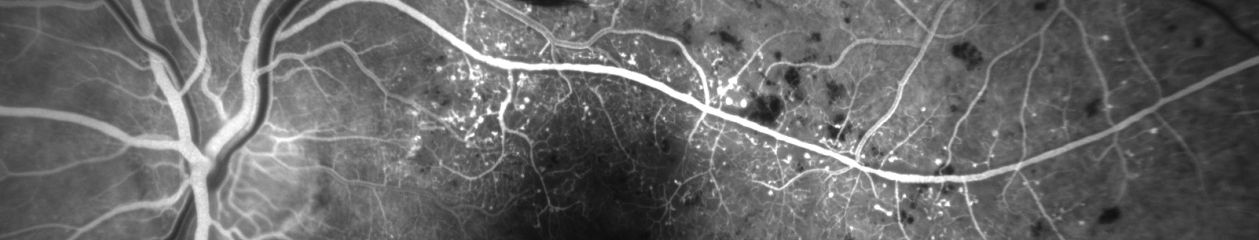
I first heard this expression many years ago when a retina specialist was explaining the results of a fluorescein angiogram to a patient and her family. He went on to explain that her retina represented the “film” in the camera and that it was swollen with fluid and wrinkled, which was the cause of her blurred vision. Everyone present easily understood the analogy, as 35mm cameras were commonplace at the time. The idea of warped or wrinkled film resonated as an analogy.    

Although it was my first time hearing this comparison, it was not a new or unique concept. The compelling connection between the eye and camera dates as far back as the 16th century when Leonardo DaVinci drew a comparison between the camera obscura and the human eye. In the next century, Della Porta, Cigoli, and Descartes also made a similar connection in their writings on optics and vision.

With the introduction of practical photographic methods in 1839, the analogy soon extended beyond the optical comparison between eye and camera obscura to include the comparison between vision and imaging. As photography and cameras quickly grew in popularity, scientists, physicians and ophthalmologists such as Helmholtz and LeConte advanced the analogy between photographic camera and the eye.​

“The further explanation of the wonderful mechanism of the eye is best brought out by a comparison with some optical instrument. We select for this purpose the photographic camera. The eye and the camera: the one a masterpiece of Nature’s, the other of man’s work.”

            Joseph LeConte

​From the earliest days of photography, several investigators sought to use the camera to document the condition of the eye. Due to the technical limitations of available photosensitive materials and the difficulty in illuminating the interior of the eye, fundus photography became the “holy grail” of medical imaging. Over the next several decades there were incremental advances in optical instrumentation and photographic processes, all moving steadily towards practical examination and photography of the eye. The most notable of these milestones was the 1851 introduction of the ophthalmoscope by Helmholtz.

Lucien Howe, one of the early pioneers in fundus photography echoed the idea of eye as camera in his 1887 article in which he described having captured the first recognizable fundus photograph:

“We know that the eye itself is a camera, and when placed behind an ophthalmoscope it has pictured upon the retina an image of an eye observed.”

Lucien Howe

The analogy was taken yet a step further by Mann who presented a paper utilizing animal eyes as an actual camera by placing photographic film against an aperture cut in the posterior pole of the globe.

“The main object has been to demonstrate that the eye can actually be used as a camera, a fact which is of interest chiefly because of the frequent comparison between the two.”

William Mann


From that point on there were several milestones in the evolution of both photography and ophthalmic photography that culminated in reflex-free fundus cameras equipped with electronic flash by the 1950’s. The modern fundus camera revolutionized ophthalmic photography and provided the platform for the important development of fluorescein angiography techniques by Novotny and Alvis in 1959. Optical coherence tomography has further revolutionized ophthalmic imaging, but fundus photography and angiography remain vital tools in the diagnosis and treatment of ocular disease.

In today’s wireless and digital age, the analogy of eye as camera may not be as universally accepted as it was a generation ago. Most 35mm cameras had a spring-loaded pressure plate on the film door to ensure the film was held flat and firmly in place. It was easy to envision the concept of a piece of film (or a retina) being wrinkled, torn, or warped.

Today’s digital cameras do not open to reveal the fixed sensor inside so the analogy doesn’t resonate as well. Yet the optical comparison between the eye and camera lens still rings true today, even with sophisticated scanning instruments such as SLO and OCT.

“The eye is like a camera.”…. Since the days of DaVinci, this idea has been echoed countless times by philosophers, artists, ophthalmologists and photographers. As an ophthalmic photographer, I consider myself fortunate to have spent a career photographing the human eye: one camera being used to preserve the capabilities of the other.

“The camera exists because men were intrigued by the function of the eye and wished to be able to reproduce on a permanent record that which the eye enabled the brain to record. How appropriate then that ophthalmology has turned the camera into a valuable tool for recording the structures of the eye.”

R. Hurtes

“The eye is like a camera.”

References

Wade NJ, Finger S. The eye as optical instrument: from camera obscura to Helmholtz’s perspective. Perception 2001; 30:1157-1177

LeConte J. The Eye as an Optical Instrument. from Sight: An Exposition of the Principles of Monocular and Binocular Vision. 1897

Howe L. Photography of the interior of the eye. Trans Amer Ophth Soc. 23:568-71 July 1887

Mann WA. Direct utilization of the eye as a camera. Trans Am Ophthalmol Soc 42:495-508, 1944

Novotny HR, Alvis DL. A method of photographing fluorescence in circulating blood of the human retina. Circulation. 24:82, 1961

Meyer-Schwickerath, G. Ophthalmology and photography. AJO 66:1011, 1968

Bennett TJ. Ophthalmic imaging – an overview and current state of the art. Journal of Biocommunication, 32(2):16-26, 2007.

Bennett TJ. Ophthalmic imaging – an overview and current state of the art, part II. Journal of Biocommunication, 33(1):3-10, 2007.

Hurtes R. Evolution of Ophthalmic Photography. International Ophthalmology Clinics. 1976; 16(2):1-22