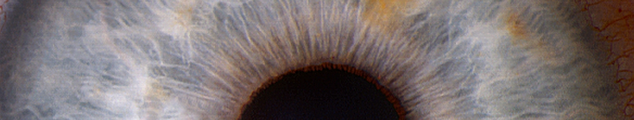
Monochromatic photography has a long history of use in ophthalmology. Illumination of the subject eye with light of a specific color can enhance the contrast and visibility of various structures or findings. Traditionally, it is used in black-and white fundus photography to enhance anatomical details of the retina and choroid, but the same concept can also be applied to other parts of the eye.
Classic example of monochromatic green (red-free) rendition of fundus pathology. The green filter enhances the view to the retinal vessels and the macular lesion.
Monochromatic information can be captured by either filtering the light source (such as with a fundus camera), or placing a contrast filter in front of the camera lens to limit the color reaching the sensor. A subject color will appear lighter when photographed through a filter of the same color, and darker when photographed through a filter of its’ complementary or opposite color. For example, a red subject would appear lighter if exposed through a red filter and darker when photographed a complementary color filter, which in this case would be cyan (blue-green).
Red/Green/Blue color separations of a color fundus photograph of a choroidal nevus demonstrating the value on monochromatic rendering. The red channel shows a darkened lesion similar to the monochromatic effect using a red contrast filter. The blue and green channels suppress the view of the pigmented lesion.
In addition to the traditional technique of using monochromatic illumination with black-and-white photography, another alternative is to take full-color photos without filters and then use software to split the full color image into separate red, green, and blue color components.
Monochromatic views of a salmon colored conjunctival lesion. The lesion appears lighter than the conjunctiva in the red channel, while the blues channel darkens the lesion making it more apparent.A similar effect occurs in this image of subconjunctival hemorrhage. It’s a great example of how subject colors that are the same as the channel or filter will appear lighter. The blood is darkened by the green and blue channel.
This is a remarkably simple way to obtain monochromatic renderings from any full color image. It works particularly well with color slit-lamp photos of the anterior segment.
A conjunctival lesion stained with lissamine green. Here the red channel darkens the stain pattern, while the green and blue channel lightens the blue-green dye.
One disadvantage to this method is the loss of resolution that occurs when viewing just a single channel that makes up the full color image. It also limits the available monochromatic information to just the three primary colors, red, green, and blue, but that’s usually sufficient for anterior segment applications.
Monochromatic renditions of corneal blood staining.A dislocated cataract in the anterior chamber. Blue (which is the opposite color of yellow) darkens the lens almost completely, while the red channel enhances its appearance.
When performing fundus photography or angiography, patients often ask about the technology used to help diagnose their ocular condition. After explaining that it’s a form of photography, the conversation will often turn to cameras. Patients sometimes ask for advice on what type of camera to buy for personal use. Often they’ll tell me about a particular camera they have and will invariably say “the camera takes great pictures”.
I’ve always found this expression and the concept behind it quite amusing. It assumes the person looking through the viewfinder and pressing the shutter button has nothing to do with it! On several occasions these conversations took place at a VA hospital while photographing vets. They’ll talk about having purchased a camera overseas, such as a Voigtlander, Zeiss Contax, or Leica in Europe during WWII, or a Nikon while stationed in Japan, Korea, or Vietnam. Without fail, they’ll tell me that “the camera takes great pictures”. All are quality cameras with good optics, but none of them are capable of taking pictures on their own. Someone has to compose the image and press the shutter.
A related misconception has occurred in ophthalmic imaging over the past decade. With all the automated features such as eye tracking, sampling, auto-alignment and auto-exposure, etc. in the current crop of instruments, there is a perception that retinal imaging is simple and easy to perform. The implication is that the machine takes the picture, not the operator.
It’s true that recent advancements in technology have improved and simplified image capture, but I still believe that skilled photographers produce the best diagnostic images. I once had a conversation with an overly-enthusiastic OCT salesman who claimed his instrument was better than the competition. As proof, he showed me two images of the same patient taken with two different OCT instruments. “See”, he said, “The competition’s instrument missed the pathology”. I corrected him saying that it wasn’t the instrument that missed the pathology, but the operator.
High resolution line scan of a small retinal arterial macroaneurysm captured using the free scanning technique.
This sparked an interesting debate. I argued that a skilled OCT operator, given appropriate direction from the ordering physician, would not have missed the area of interest. Ultimately we agreed it’s up to the operator to make the best use of the technology they have available.
Several famous photographers have weighed in on the relationship between camera and photographer in producing great images. I love these quotes:
“You don’t take a photograph, you make it.” Ansel Adams
“The camera doesn’t make a bit of difference. All of them can record what you are seeing. But, you have to SEE”. Ernst Haas
“The camera is an instrument that teaches people how to see without a camera.” Dorothea Lange
“Photography is the art of observation… I have found it has little to do with the things you see and everything to do with the way you see them.” Elliot Erwitt
“There is nothing worse than a brilliant image of a fuzzy concept.” Ansel Adams
And finally, whenever someone states, “This camera takes great pictures”, I reply with another common quote that’s been attributed to several people, including boxing trainer Roger Mayweather, basketball great Charles Barkley, and possibly even Confucius:
“It ain’t the tools, it’s the carpenter”.
Slit-lamp photograph of dislocated intraocular lens implant and iris erosion shown in transillumination. Slit-lamp imaging relies on advanced photographic lighting techniques to accurately document the condition of the eye.
Voigtlander camera image from Wikimedia Commons: https://en.wikipedia.org/wiki/File:Vitorets.JPG#filelinks