
Although it may seem like a recent phenomenon, the photographic self-portrait has been with us since the dawn of photography. Perhaps the earliest known “selfie” was taken by Hippolyte Bayard (Portrait of a Drowned Man), a Frenchman who claimed to have invented a photographic process prior to the Daguerrotype. The same can be said of William Henry Fox-Talbot (The Reading Establishment).
In recent years, the photographic self-portrait has exploded in popularity into a global phenomenon, fueled by social media sites such as Facebook, Instagram, Twitter, Snapchat, and others. It is estimated that over one million “selfies” are taken every day. A recent search of Instagram returned over 211 million photos with the hashtag “#selfie”. The term “selfie” is believed to have originated in Australia and has been elevated from internet slang to our common vernacular and even inclusion in several formal English dictionaries. In fact, selfie was Oxford Dictionary’s word of the year for 2013!
Everybody seems to be getting into the act including celebrities, politicians, and even the Pope! Selfies are even popular in space. Astronauts have shared several spacewalk selfies online, and last year, NASA promoted a global selfie project to celebrate Earth Day. They solicited over 36,000 selfies from around the world and created an interactive composite image that can be viewed on their website.
The selfie is so ubiquitous in today’s pop culture that cell phones and digital cameras often include built-in selfie-friendly apps and features such as extra wide angle lenses, articulating screens or front facing screens that facilitate the selfie pose. You can also purchase selfie sticks to extend the camera to a better vantage point. These popular items go well beyond the simple self-timer found on many cameras of yesteryear. Selfies are often purposely self-deprecating, campy, cheesy, or irreverent. They are meant to be spontaneous and fun and are not usually taken very seriously. The selfie craze has even spawned the infamous “duck face” pose.
Despite the fun and seemingly harmless spirit behind them, there is a belief that taking selfies can be a sign of narcissism rather than simple self-expression. There is also some concern it can be addicting and unhealthy. But there is a growing trend in telemedicine where patients can take and forward selfies to their doctors to help diagnose or triage the urgency of their condition. So maybe there are some legitimate uses for selfies.

As a life-long photographer, I’ve taken my share of selfies over the years. I’ve even attempted a few with the equipment I use for diagnostic ophthalmic photography. Some are goofy, and in the spirit of social media selfies. Some are more practical.
Most ophthalmic imaging devices are not what you’d normally consider selfie friendly, at least not in terms of taking a photo of one’s own eyes. Because the optics of these devices are designed for photographing curved surfaces found in the interior of the eye, they usually create distortion when backed up an appropriate distance to take a facial portrait. The effect of this distortion eliminates the need for a goofy facial expression if your goal is just to post a unique selfie on the internet!

But what about useful diagnostic or artistic photos of your own eyes, photos that go beyond distorted face selfies? It’s not only possible, but surprisingly good images can be obtained with some devices. Non-mydriatic instruments with a monitor that can be pivoted toward the patient/photographer lend themselves to self-imaging, while those with an optical viewfinder (fundus camera) or fixed monitor position (Cirrus) do not. I’ve been able to obtain eye selfies with the Zeiss Stratus, Heidelberg HRT, Heidelberg HRA/OCT, Clarity RetCam, Tomey specular microscope and various handheld external cameras. But, you might ask, “So what?” or “Why?”

Well, there have been times when I needed to check a device during maintenance or a software upgrade and it was convenient to use myself as the patient. Sometimes while training staff to use a device, I’ll demonstrate the procedure on myself. Other times, I’ve needed a quick example of a “normal” eye for a lecture like those above.

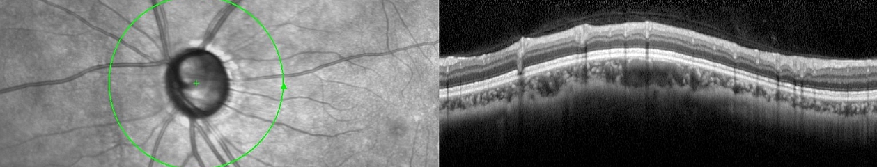
Surprisingly, the ability to take eye “selfies” has helped me identify and track pathology in my own eyes. Two years ago I suffered an idiopathic retinal tear with avulsed bridging vessel and persistent vitreous hemorrhage. This was successfully treated with vitrectomy. Like many patients, I developed a cataract after the vitrectomy. I also began to notice some distortion that corresponded to progression of an epiretinal membrane (ERM) in the same eye.

Any time I noticed a change in vision I would repeat an OCT on myself. Over the course of six months I tracked an increase in thickness of about 100 microns. The cataract also progressed and I was scheduled for cataract surgery. Two weeks prior to surgery I noticed a very subtle change in vision and sat down at the OCT like I’d done several times in the past. The OCT detected some cystoid macular edema (CME) from the ERM. Picking up the CME prior to cataract surgery was very beneficial. Preexisting CME can be exacerbated by cataract surgery, so my surgeon began a course of treatment that reduced the edema. My OCT selfies likely helped us avoid more severe or persistent edema by catching it in advance.
Cataract surgery went as planned, but within a few hours of my procedure I began to notice a new visual abnormality: a paracentral gray scotoma. Upon arriving at the clinic the next day for my post-operative check, I immediately did an SLO/OCT selfie and identified an unusual finding that corresponded directly to the scotoma.

SD-OCT demonstrated an area of hyper-reflectivity in the middle retinal layers just temporal to the fovea (green arrows) and the IR reflectance image showed a distinct dark gray lesion. Fortunately, the scotoma began to fade within a few days and so did the lesion. The jury is still out on the exact cause of the lesion but the selfies have enabled us to track improvement of my condition and possibly publish a case report. We believe it may be a case of paracentral acute middle maculopathy (PAMM), a recently described variant of acute macular neuroretinopathy (AMN). It’s rare enough, that I was able to present it at the OPS Rare Case Symposium in Ann Arbor.

As you can see, image quality can be quite good with a little practice. So good in fact that I’ve received a bill from my institution for OCT images that I’ve performed on myself! Here is a double selfie video of a Spectralis IR fundus image showing how easy it is to capture my own epiretinal membrane.
“
I’m beginning to think maybe I should stop taking selfies of my own eyes. After all I keep finding abnormalities! But there is a growing trend in telemedicine where patients can take and forward selfies to their doctors to help diagnose or triage the urgency of their condition.
Ophthalmic Photographers taking diagnostic selfies: obsessive, silly, or beneficial?