Tag Archives: selfie

Celestial Bodies – The Eye and Space

First time viewers of ophthalmic images frequently make the observation that the photos look like something from outer space. Especially when reviewing the round orange retinal photos with their eye doctor, patients often comment, “That looks like the planet Mars.”

Every time it happens I get a chuckle out of it. As if we all truly know what the planet Mars really looks like! But to most people, images of the inside of an eye are foreign and amazing. And there does seem to be a little science fiction aspect to both the appearance of the eye when viewed at high magnification, as well as the technology used to capture these amazing images. There are however, several space analogies that really seem to ring true. Among eye-care professionals, the eyeball is routinely referred to as the “globe”.

Macular Star in a patient with cat scratch neuroretinitis

Many clinical findings are named by their appearance rather than an underlying cause, and several conditions have names derived from their similarity in appearance to objects in space: asteroid hyalosis, macular star, star folds, starry sky, astrocytoma, stellate pattern, etc.

Asteroid hyalosis is comprised of calcium soaps suspended in the vitreous cavity behind the iris. Despite their appearance, most patients with this condition are asymptomatic.

In fact, there are enough conditions like this, that I’ve been able to compile them into the Ophthalmic Jeopardy category: Celestial Bodies.

Transillumination of a thinly pigmented iris in a patient with ocular albinism.

Like images from space, there does seem to be an element of wonder and mystery when we peer inside the globe, so in some ways the analogy makes sense.
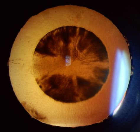
Cataract

Many ophthalmic images seem reminiscent of photographs from NASA. Or they may stir our imagination or perception of how objects in space might appear.

Lisch nodules on the iris of this patient with neurofibromatosis are reminiscent of peaks, valleys and craters seen in NASA photographs from planets or moons in our solar system.

There are other connections as well. Some of the photographic techniques used by both astronomers and ophthalmic photographers are actually similar. IR capture, interferometry and stereo imaging are common techniques in both fields.  The principles of  rotational stereo imaging can be applied to both subjects. Filters or lasers of different wavelengths are commonly used to enhance visibility of certain features in both subject types.

Most of these analogies between the eye and outer space, are loose associations rather than a direct connection. There is however, at least one eye condition that can be directly associated with a celestial body. Solar retinopathy is a type of photic injury to the retina that is the result of staring at the sun. This condition typically occurs in patients with psychiatric disorders or under the influence of hallucinogenic drugs.

A case of solar retinopathy with a subtle yellow-white foveal lesion with associated early pigmentary changes.

Some scholars believe that early astronomers, especially Gallileo, went blind as the result of solar retinopathy from viewing the sun through a telescope. It’s important to note that this condition can also occur from viewing a solar eclipse without protective eyewear. The upcoming solar eclipse visible in the U.S. on August 21, may cause a spike in cases of solar retinopathy presenting to emergency rooms and eye clinics. The American Academy of Ophthalmology offers some tips for safe viewing of the eclipse.

In recent years, another connection between outer space and vision has been discovered. It turns out that space travel can have some damaging effects on the human eye. Long-term exposure to microgravity can lead to a hyperopic shift in vision from flattening of the globe. This condition is believed to be related to increased intracranial pressure and is sometimes associated with optic disc edema, cotton wool spots and choroidal folds. Optical coherence tomography (OCT) is used to document  changes in thickness of the retinal nerve fiber layer of astronauts before, during, and after space flight.

I took this OCT selfie a few years back when we had a scientist from NASA visiting our clinic while exploring the possibility of putting an OCT on the International Space Station. She wanted to see the Heidelberg Spectralis in clinical use. After demonstrating on several patients, the scientist asked me if I thought it were possible for someone to take an OCT image of themselves. I pivoted the monitor, control panel, and footswitch around so I could operate the OCT from the patient chair and then captured some images of my own retina. I was showing off a little and smugly cautioned the NASA doctor that this was a difficult feat that only an experienced ophthalmic imager could perform. After all, I’ve been doing this for over thirty years. She paused for a moment and then said, “With all due respect, astronauts are some of the smartest and most talented people on earth. They shouldn’t have any difficulty performing OCTs on themselves after a some brief training.” Suddenly I didn’t feel so smug.

A year or so later, the Spectralis arrived at the International Space Station and it looks like she was right. I heard from some colleagues at Heidelberg that the astronauts were given less than 30 minutes of training on the instrument and mastered it quickly!

It’s pretty cool knowing that astronauts are performing ophthalmic imaging on the International Space Station. I wonder if they ever see any resemblance between the eye and celestial bodies?

Disclosure: I have no financial or proprietary interest in the Heidelberg Spectralis.

Here are some links on the condition that’s effecting the vision of astronauts and the use of diagnostic imaging on the space station:

http://www.vision-research.eu/index.php?id=858

https://www.theatlantic.com/science/archive/2017/01/seeing-in-space/513650/

http://www.utsouthwestern.edu/newsroom/news-releases/year-2017/jan/vision-levine.html

https://www.washingtonpost.com/news/speaking-of-science/wp/2016/02/11/cosmic-breakthrough-physicists-detect-gravitational-waves-from-violent-black-hole-merger/

https://www.healio.com/ophthalmology/retina-vitreous/news/print/ocular-surgery-news/%7B5cd3865b-b1db-473a-8800-e3e3b50c6df5%7D/monitoring-long-term-effects-on-vision-in-microgravity-a-priority-for-nasas-future-flight-to-mars


ICOP – Singapore

I’m still feeling a little jet lagged after traveling halfway around the world, but what an amazing trip! Along with over seventy other ophthalmic imagers, technicians, and physicians, I was in Singapore to attend the 2017 International Conference on Ophthalmic Photography (ICOP).

ICOP is a joint educational venture between several ophthalmic imaging organizations including the Ophthalmic Photographers’ Society (OPS) from the United States, the Ophthalmic Imaging Association (OIA) from the UK, the Australian Institute of Medical and Biological Illustrations (AIMBI) from Australia, and the Ooghelkundige Fotografie Nederland (OFN) from the Netherlands. Delegates from 15 different countries were in attendance at this conference.

The three day program was held at the Singapore National Eye Centre (SNEC).  The educational program put together by Paula Morris, CRA, FOPS and Sarah Armstrong, CRA, OCT-C, FOPS, included invited lectures, special keynote lectures, and Scientific Paper sessions from dlegates in attendance. Keynote lecturers included Wong Tien Yin, MD, PhD discussing: How a Fundus Photograph Can Save Your Life, Giovanni Staurenghi, MD who presented: Old and New Angiography, Suber Huang, MD who showed amazing images in his lecture: The ASRS Image Bank – a Worldwide Legacy and Gavin Tan Discussing: OCT Angiography – Changing the Way We See.

Photo by Chris Barry, FOPS

I was honored to attend ICOP as not only a delegate, but as an invited lecturer. I ended up presenting all three days of the conference and it was a great honor for me to contribute to the program in this way. I chose topics that I felt would appeal to an international audience and I think it worked out okay. I presented a version of Ophthalmic Jeopardy! that I customized for an audience that was unfamiliar with the namesake television quiz show, that while famous in the U.S., isn’t broadcast in Singapore. Talk about performing a high wire act without a net! I made sure I had some content that everyone could relate to including local Singapore trivia and a review of content covered by presenters in many of the earlier lectures.

The imaging staff at SNEC is renowned for the quality of their ophthalmic photography and they were clearly happy to be hosting this event on their home turf. On a tour of the facility, award winning images were displayed prominently on the walls of the imaging department. It was inspiring to see such an amazing collection of work of the highest quality. Photographers Joseph Ho, Kasi Sandhanam and the rest of the SNEC staff are amazing imagers that are able to balance the efficiency needed to handle a high volume of patients with the highest standards in image quality. They are true professionals in our field.

John Leo with his award winning image. Photo by Chris Barry, FOPS.

Speaking of high quality imaging, the conference also included a photo competition and exhibit that showed some incredible clinical and artistic imaging. Award winners included Sarah Armstrong, Lisa Brealey, Angela Chappell and John Leo.

Photo by Chris Barry, FOPS.

In addition to the educational content during the conference, there were exhibits by a number of sponsoring vendors, some incredible refreshments during the breaks, and a fun evening of food, music, and comradery at the welcome reception.

A highlight of the reception was the photo booth that not only produced mementos of the occasion, but acted as the perfect icebreaker, as spontaneous groups of old friends and new acquaintances would pose together in the spirit of ICOP!

Of course as professional imagers, most attendees had cameras with them and spontaneous selfies were popping up everywhere!

This is the second time that ICOP has been held in this thoroughly modern and spectacular city of Singapore, having previously been hosted here in 1990. And what a great venue for an international conference! Singapore represents an incredible blend of Asian cultures, British influence, modern architecture and great weather.

Like many other delegates I tried to visit as many of the popular sightseeing spots as possible including, Marina Bay, Merlion Park, Super Trees, Sentosa Island, Chinatown, Buddha Tooth Temple, Hawker Markets, the Mt. Faber  Cable Car, Henderson Waves, Botanical Gardens and many more. With all these famous sights and numerous museums, there is so much to do and see in this amazing city.

Local residents Paul Chua and Albert Sim took some time to show us some of the local sights in the evenings and recommend the best food stalls in the hawker markets. Joseph Ho hosted an amazing dinner of chili crab at Jumbo Seafood. Alan Wee wrote a great blog post for the OPS/ICOP website with a list and map of places to visit, along with suggestions for some of the best food and coffee shops in the city. It was great having such knowledgeable local guides to help us experience all that Singapore has to offer.

Like many other ICOP delegates, I tried to take in as many of these sights as possible. One of the attractions on my list was the Trick Eye Museum on Sentosa Island which seemed like something an eye imaging professional should check out, at least for a laugh or two. It’s a place where you can take some really cheesy selfies with props and silly scenes in the background! Although I didn’t have time to visit, I walked past and snapped a photo or two. Maybe next time.

ICOP 2017 was an amazing success. Kudos to the international ICOP planning team of Paula Morris, Sarah Armstrong, Chris Barry, Ethan Priel, Becky MacPhee, Angela Chappell and Gerard de Graaf.

The SNEC staff and organizing committee were incredible hosts from Gemmy Cheung, MD, Wong Tien Yin, MD, Gavin Tan, MD,  Dr Thiyagarajan Jayabaskar and Lim Hui San, to Joseph Ho, Kasi Sandhanam, and the rest of the imaging and AV teams. They really know how to put on a professional conference.

It was great seeing old friends from around the globe as well as make several new ones.

Photo by Chris Barry, FOPS

ICOP promotes networking with colleagues and seems to make the world just a little smaller. I believe that each of us found that we all have so much in common no matter how far apart we live. I look forward to the next ICOP which will take place in 2020 at a location yet to be determined.

*ICOP group photo by Govindarjan Jayaraman.


Me, Myself, and Eye

Although it may seem like a recent phenomenon, the photographic self-portrait has been with us since the dawn of photography. Perhaps the earliest known “selfie” was taken by Hippolyte Bayard (Portrait of a Drowned Man), a Frenchman who claimed to have invented a photographic process prior to the Daguerrotype. The same can be said of William Henry Fox-Talbot (The Reading Establishment).

In recent years, the photographic self-portrait has exploded in popularity into a global phenomenon, fueled by social media sites such as Facebook, Instagram, Twitter, Snapchat, and others. It is estimated that over one million “selfies” are taken every day. A recent search of Instagram returned over 211 million photos with the hashtag “#selfie”. The term “selfie” is believed to have originated in Australia and has been elevated from internet slang to our common vernacular and even inclusion in several formal English dictionaries. In fact, selfie was Oxford Dictionary’s word of the year for 2013!

Everybody seems to be getting into the act including celebrities, politicians, and even the Pope! Selfies are even popular in space.  Astronauts have shared several spacewalk selfies online, and last year, NASA promoted a global selfie project to celebrate Earth Day. They solicited over 36,000 selfies from around the world and created an interactive composite image that can be viewed on their website.

The selfie is so ubiquitous in today’s pop culture that cell phones and digital cameras often include built-in selfie-friendly apps and features such as extra wide angle lenses, articulating screens or front facing screens that facilitate the selfie pose.  You can also purchase selfie sticks to extend the camera to a better vantage point.  These popular items go well beyond the simple self-timer found on many cameras of yesteryear. Selfies are often purposely self-deprecating, campy, cheesy, or irreverent. They are meant to be spontaneous and fun and are not usually taken very seriously. The selfie craze has even spawned the infamous “duck face” pose.

Despite the fun and seemingly harmless spirit behind them, there is a belief that taking selfies can be a sign of narcissism rather than simple self-expression. There is also some concern it can be addicting and unhealthy. But there is a growing trend in telemedicine where patients can take and forward selfies to their doctors to help diagnose or triage the urgency of their condition.  So maybe there are some legitimate uses for selfies.

As a life-long photographer, I’ve taken my share of selfies over the years. I’ve even attempted a few with the equipment I use for diagnostic ophthalmic photography. Some are goofy, and in the spirit of social media selfies. Some are more practical.

Most ophthalmic imaging devices are not what you’d normally consider selfie friendly, at least not in terms of taking a photo of one’s own eyes. Because the optics of these devices are designed for photographing curved surfaces found in the interior of the eye, they usually create distortion when backed up an appropriate distance to take a facial portrait. The effect of this distortion eliminates the need for a goofy facial expression if your goal is just to post a unique selfie on the internet!

But what about useful diagnostic or artistic photos of your own eyes, photos that go beyond distorted face selfies? It’s not only possible, but surprisingly good images can be obtained with some devices.  Non-mydriatic instruments with a monitor that can be pivoted toward the patient/photographer lend themselves to self-imaging, while those with an optical viewfinder (fundus camera) or fixed monitor position (Cirrus) do not. I’ve been able to obtain eye selfies with the Zeiss Stratus, Heidelberg HRT, Heidelberg HRA/OCT, Clarity RetCam, Tomey specular microscope and various handheld external cameras. But, you might ask, “So what?” or “Why?”

Well, there have been times when I needed to check a device during maintenance or a software upgrade and it was convenient to use myself as the patient. Sometimes while training staff to use a device, I’ll demonstrate the procedure on myself. Other times, I’ve needed a quick example of a “normal” eye for a lecture like those above.

Surprisingly, the ability to take eye “selfies” has helped me identify and track pathology in my own eyes. Two years ago I suffered an idiopathic retinal tear with avulsed bridging vessel and persistent vitreous hemorrhage. This was successfully treated with vitrectomy. Like many patients, I developed a cataract after the vitrectomy. I also began to notice some distortion that corresponded to progression of an epiretinal membrane (ERM) in the same eye.

Any time I noticed a change in vision I would repeat an OCT on myself. Over the course of six months I tracked an increase in thickness of about 100 microns. The cataract also progressed and I was scheduled for cataract surgery. Two weeks prior to surgery I noticed a very subtle change in vision and sat down at the OCT like I’d done several times in the past. The OCT detected some cystoid macular edema (CME) from the ERM. Picking up the CME prior to cataract surgery was very beneficial. Preexisting CME can be exacerbated by cataract surgery, so my surgeon began a course of treatment that reduced the edema. My OCT selfies likely helped us avoid more severe or persistent edema by catching it in advance.

Cataract surgery went as planned, but within a few hours of my procedure I began to notice a new visual abnormality: a paracentral gray scotoma. Upon arriving at the clinic the next day for my post-operative check, I immediately did an SLO/OCT selfie and identified an unusual finding that corresponded directly to the scotoma.

SD-OCT demonstrated an area of hyper-reflectivity in the middle retinal layers just temporal to the fovea (green arrows) and the IR reflectance image showed a distinct dark gray lesion. Fortunately, the scotoma began to fade within a few days and so did the lesion. The jury is still out on the exact cause of the lesion but the selfies have enabled us to track improvement of my condition and possibly publish a case report. We believe it may be a case of paracentral acute middle maculopathy (PAMM), a recently described variant of acute macular neuroretinopathy (AMN). It’s rare enough, that I was able to present it at the OPS Rare Case Symposium in Ann Arbor.

As you can see, image quality can be quite good with a little practice. So good in fact that I’ve received a bill from my institution for OCT images that I’ve performed on myself! Here is a double selfie video of a Spectralis IR fundus image showing how easy it is to capture my own epiretinal membrane.

I’m beginning to think maybe I should stop taking selfies of my own eyes. After all I keep finding abnormalities! But there is a growing trend in telemedicine where patients can take and forward selfies to their doctors to help diagnose or triage the urgency of their condition.

Ophthalmic Photographers taking diagnostic selfies: obsessive, silly, or beneficial?