One of the best ways to ensure high quality ophthalmic diagnostic images is to first establish a rapport with the patient. Once you’ve gained their trust you can easily guide them through the sometimes uncomfortable or stressful process of obtaining information that may determine their diagnosis and influence treatment decisions. Chatting them up a little usually helps to ease their nervousness. This friendly interaction often leads to some interesting conversations.
Patients who undergo ophthalmic imaging for the first time are especially amazed by the whole experience. They are fascinated by the technology used and the incredible images we are able to obtain.
But they also want to know a little about the qualifications needed to perform these tests. They may ask questions like, “What is your job called?” Or, “How long have you been doing this?” “Do you need to be a doctor to do this?” But the most common question I get is, “How did you get into this field?” I usually chuckle and then give them my stock answer, “I’m still trying to figure out how that happened!” I go on to explain it’s not something I set out to do as a profession.
Their next comment is usually, “You must have gone to school a long time, or have a special degree, in order to do this.” They are shocked when I tell them that most people in the field were trained on the job and that there aren’t any specific educational or licensure requirements. It seems to put them at ease when I tell them that I hold multiple certifications in ophthalmic imaging. This speaks to the value of achieving and maintaining voluntary certification.
So how does one get started in this field?
There’s no one “right way” to become an ophthalmic imager. In the absence of formal training or degrees in our profession, most of us learn the majority of the required skills on the job. My background was in commercial photography and I gradually learned enough ophthalmology to apply my photographic skills to imaging the eye. There are many others in the field with a background in photography. Others started as medical assistants or technicians and then learned the photographic side of things on the job. The point is that very few of us set out to become an ophthalmic photographer and one day find ourselves working in the field and learning many of the requisite skills on the job.
One of the problems with OJT is that it is often limited to what is being done in that particular practice setting. To get started in the field or obtain more comprehensive training and expand your skills you may want to take a look at some currently available resources such as text books, online tutorials here on eye-pix.com as well as educational resources through these sources:
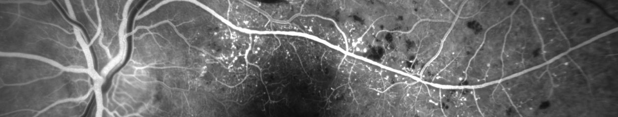
Ophthalmic imaging plays a vital role in the documentation and diagnosis, of a wide variety of ocular diseases. It is a fascinating profession, yet obtaining the requisite knowledge and skills to perform diagnostic imaging at a high level can be a challenge. Some of the fundamental techniques and technology used to image the eye have remained the same for many years, but there have been several major advances in the profession over the last two decades. Spectral Domain OCT, fundus autofluorescence, multi-modal imaging, scanning laser ophthalmoscopes, OCT angiography and other techniques have altered the imaging landscape and require that even experienced imagers learn new skills.
There is very little formal education available in ophthalmic imaging, with the notable exception of the ophthalmic imaging curriculum offered at the Rochester Institute of Technology. Graduates of the program at RIT represent some of the most prepared and successful professionals in our field. The vast majority of practitioners in our profession however, do not have this educational foundation. Most of us find it necessary to learn some of the required knowledge and skills through other methods, such as seminars, workshops, handbooks, instrument manuals, and online resources. These are all valuable educational tools, but typically don’t provide a complete educational foundation.
We now have a new resource to help us learn and stay up to date. Ophthalmic Imaging: Posterior Segment Imaging, Anterior Eye Photography and Slit Lamp Biomicrography by Professor Christye Sisson, is an important new educational resource for both novice and experienced imagers. It is the first comprehensive textbook in ophthalmic imaging published in over fifteen years. Our profession really needs a resource such as this, and Professor Sisson is uniquely positioned to create such an educational text. After several years as a clinical ophthalmic photographer, she transitioned into a career as an educator in photography and ophthalmic imaging. She currently serves Program Chair of Photographic Sciences at RIT and this text is based on the curriculum she developed for her students there.
Textbooks can be somewhat analogous to teachers. To be effective they need to be organized, accurate, stimulating, and easy to understand. The organization and layout of this book recognizes the varied career paths for entry into the profession. This text covers fundamentals such as ocular anatomy and photo technology for those just getting started in the field, as well as new technologies that are now dominating our profession. It is exciting to know we have a new “teacher” available to learn the basics of ophthalmic imaging, build new skills, or prepare for professional certification.
I can’t wait to get a look at the final product.
Update 7/1/2018: I received my copy of this text a couple months back and have had time to delve into it more deeply. It is everything that I expected it to be and more. It’s been a useful reference when I wanted to quickly look up topics such as ultra wide-field imaging and scanning laser ophthalmoscopy. I’m sure I’ll continue to reach for it whenever I need a reference in ophthalmic imaging.
The act of learning through a medium that both educates and entertains.
Any of various media, such as computer software, that educate and entertain.
When I am invited to speak at educational meetings, one of the most requested and popular presentation topics is a program titled, Ophthalmic Jeopardy! Based on the popular television quiz show format that most everyone is familiar with, I’ve created an interactive learning experience that also manages to entertain. In short, it’s “Edutainment”. It’s not a new or novel idea, but I’ve taken it a step or two further than similar game show presentations in ophthalmic education. The evolution of Ophthalmic Jeopardy! is interesting.
Years ago, one of the faculty members at the Penn State Department of Ophthalmology approached me about improving our local technician education program. The program included a simple quiz-show format of questions-and-answers with the host reading questions out loud from hand-written cards. It worked, but he wanted to “jazz things up a little”. He told me he had done some online research and found a source for Jeopardy style lockout buzzers/lights that would allow contestants to buzz in when they knew the correct answer. He wanted to pick contestants from the audience and turn it into a competition. Now all he needed was a way to project the questions onscreen and asked if we could make it more interactive like Jeopardy, with onscreen columns of different question-and-answer categories.
I gave it some thought and told him it was possible, but entirely too much work to warrant the effort. But he knew me too well! I gave it a little more thought and started tinkering with the use of hyperlinks in PowerPoint to build an interactive screen that would allow us to randomly move back and forth between categories and questions. I had attended an OPS course entitled “Whiz-Bang PowerPoint Presentations” where Bill Anderson shared a way to hyperlink menus to organize an educational program with easy navigation between multiple speaker presentations. I figured I could build a Jeopardy template using similar hyperlinks between slides.
I converted our existing quiz questions into the Jeopardy answer and question format, but many of them were simple or dry examples. Taking inspiration from Jeopardy! and the sometimes tongue-in-cheek themes, I began accumulating new questions and categories that would entertain as well as test knowledge. In crafting questions, I’ve relied on many years of training and experience in writing questions for certification examinations. But instead of being restricted by the necessary rules for crafting certification questions, Jeopardy allowed me to have some fun and take liberties with some of the topics and content.
Suddenly the project grew and seemed to take on a life of its own. Each presentation contains over 250 hyperlinks, tons of photos, videos, and sound files. We can now chose from an ever growing bank of questions that numbers in the several hundreds! The content is never the same twice. Each time I present it, there are new content areas and questions, but I keep some of the core categories. Often the content will be customized to the specific audience; for example including a local trivia category at regional or international meetings. It’s a great way to review content from other presentations over the course of a day-long or multi day meeting.
In the early days, we would pick contestants who would use the buzzers to buzz in when they knew the answers, we kept score, and gave prizes to the winners. Different faculty members from Penn State Ophthalmology acted as the host and relished playing the part of Alex Trebek. I was the “puppet master” behind the scenes, driving the program and selecting the appropriate hyperlinks to navigate through the questions.
Something was still missing however. It was an entertaining spectacle, but the majority of the audience was reduced to bystanders when we could only chose five contestants from the group. So we eventually opened it up to the entire audience rather than a handful of contestants. At times it can become a little chaotic this way, but everyone seems engaged and involved.
Although we’ve used it at Penn State for audiences ranging from physicians, technicians and the general public, the version used at photography meetings has a higher level of both difficulty and “cheesiness”. Imagers seem to quickly recognize rare and unusual eye findings, but also have a warped sense of humor and “get” the tongue-in-cheek nature of the categories and questions. It works best with larger audiences so it’s become a staple at OPS Mid-Year Educational Programswhere the entire group is together in one lecture hall.
For the last several years, I’ve come out from “behind the curtain” and started hosting Ophthalmic Jeopardy myself. When I retire from ophthalmic photography, maybe I can be a substitute for Alex Trebek! It’s entertaining for sure, but at its core it’s also educational. It’s a fun way to both laugh and learn – in short it’s “Edutainment”!
The concept of edutainment isn’t limited to Ophthalmic Jeopardy. It seems to make it’s way into many of my presentations such as: Stereopalooza, OCT- Anatomy of a Scan, Cases That Tell a Story, Top Ten Uses of a 2×4 in Ophthalmology and others.
For information on how you can be in the audience for the next episode of Ophthalmic Jeopardy!click here.
I’m still feeling a little jet lagged after traveling halfway around the world, but what an amazing trip! Along with over seventy other ophthalmic imagers, technicians, and physicians, I was in Singapore to attend the 2017 International Conference on Ophthalmic Photography (ICOP).
ICOP is a joint educational venture between several ophthalmic imaging organizations including the Ophthalmic Photographers’ Society (OPS) from the United States, the Ophthalmic Imaging Association (OIA) from the UK,the Australian Institute of Medical and Biological Illustrations (AIMBI) from Australia, and the Ooghelkundige Fotografie Nederland (OFN) from the Netherlands. Delegates from 15 different countries were in attendance at this conference.
The three day program was held at the Singapore National Eye Centre (SNEC). The educational program put together by Paula Morris, CRA, FOPS and Sarah Armstrong, CRA, OCT-C, FOPS, included invited lectures, special keynote lectures, and Scientific Paper sessions from dlegates in attendance. Keynote lecturers included Wong Tien Yin, MD, PhD discussing: How a Fundus Photograph Can Save Your Life, Giovanni Staurenghi, MD who presented: Old and New Angiography, Suber Huang, MD who showed amazing images in his lecture: The ASRS Image Bank – a Worldwide Legacy and Gavin Tan Discussing: OCT Angiography – Changing the Way We See.
Photo by Chris Barry, FOPS
I was honored to attend ICOP as not only a delegate, but as an invited lecturer. I ended up presenting all three days of the conference and it was a great honor for me to contribute to the program in this way. I chose topics that I felt would appeal to an international audience and I think it worked out okay. I presented a version of Ophthalmic Jeopardy! that I customized for an audience that was unfamiliar with the namesake television quiz show, that while famous in the U.S., isn’t broadcast in Singapore. Talk about performing a high wire act without a net! I made sure I had some content that everyone could relate to including local Singapore trivia and a review of content covered by presenters in many of the earlier lectures.
The imaging staff at SNEC is renowned for the quality of their ophthalmic photography and they were clearly happy to be hosting this event on their home turf. On a tour of the facility, award winning images were displayed prominently on the walls of the imaging department. It was inspiring to see such an amazing collection of work of the highest quality. Photographers Joseph Ho, Kasi Sandhanam and the rest of the SNEC staff are amazing imagers that are able to balance the efficiency needed to handle a high volume of patients with the highest standards in image quality. They are true professionals in our field.
John Leo with his award winning image. Photo by Chris Barry, FOPS.
Speaking of high quality imaging, the conference also included a photo competition and exhibit that showed some incredible clinical and artistic imaging. Award winners included Sarah Armstrong, Lisa Brealey, Angela Chappell and John Leo.
Photo by Chris Barry, FOPS.
In addition to the educational content during the conference, there were exhibits by a number of sponsoring vendors, some incredible refreshments during the breaks, and a fun evening of food, music, and comradery at the welcome reception.
A highlight of the reception was the photo booth that not only produced mementos of the occasion, but acted as the perfect icebreaker, as spontaneous groups of old friends and new acquaintances would pose together in the spirit of ICOP!
Of course as professional imagers, most attendees had cameras with them and spontaneous selfies were popping up everywhere!
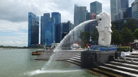
This is the second time that ICOP has been held in this thoroughly modern and spectacular city of Singapore, having previously been hosted here in 1990. And what a great venue for an international conference! Singapore represents an incredible blend of Asian cultures, British influence, modern architecture and great weather.
Like many other delegates I tried to visit as many of the popular sightseeing spots as possible including, Marina Bay, Merlion Park, Super Trees, Sentosa Island, Chinatown, Buddha Tooth Temple, Hawker Markets, the Mt. Faber Cable Car, Henderson Waves, Botanical Gardens and many more. With all these famous sights and numerous museums, there is so much to do and see in this amazing city.
Local residents Paul Chua and Albert Sim took some time to show us some of the local sights in the evenings and recommend the best food stalls in the hawker markets. Joseph Ho hosted an amazing dinner of chili crab at Jumbo Seafood. Alan Wee wrote a great blog post for the OPS/ICOP website with a list and map of places to visit, along with suggestions for some of the best food and coffee shops in the city. It was great having such knowledgeable local guides to help us experience all that Singapore has to offer.
Like many other ICOP delegates, I tried to take in as many of these sights as possible. One of the attractions on my list was the Trick Eye Museum on Sentosa Island which seemed like something an eye imaging professional should check out, at least for a laugh or two. It’s a place where you can take some really cheesy selfies with props and silly scenes in the background! Although I didn’t have time to visit, I walked past and snapped a photo or two. Maybe next time.
ICOP 2017 was an amazing success. Kudos to the international ICOP planning team of Paula Morris, Sarah Armstrong, Chris Barry, Ethan Priel, Becky MacPhee, Angela Chappell and Gerard de Graaf.
The SNEC staff and organizing committee were incredible hosts from Gemmy Cheung, MD, Wong Tien Yin, MD, Gavin Tan, MD, Dr Thiyagarajan Jayabaskar and Lim Hui San, to Joseph Ho, Kasi Sandhanam, and the rest of the imaging and AV teams. They really know how to put on a professional conference.
It was great seeing old friends from around the globe as well as make several new ones.
Photo by Chris Barry, FOPSPhoto by Chris Barry, FOPS
ICOP promotes networking with colleagues and seems to make the world just a little smaller. I believe that each of us found that we all have so much in common no matter how far apart we live. I look forward to the next ICOP which will take place in 2020 at a location yet to be determined.